Why Your Brain Has Wrinkles: New Scientific Discovery Explained
The ridges and grooves that crisscross the surface of the human brain are so familiar that we rarely stop to wonder why they exist. Yet those wrinkles—called gyri (ridges) and sulci (grooves)—are a defining feature of mammalian brains and a central clue to how our brains develop and function. Recently, scientists reported a surprising mechanism that contributes to cortical folding, reshaping a decades-old debate about whether genes, neurons, or simple physics are the chief architects of our brain’s topography. This long-form feature walks through the biology, the mechanics, and the big-picture consequences of that discovery, and explains what it means for medicine, evolution, and the study of the mind.
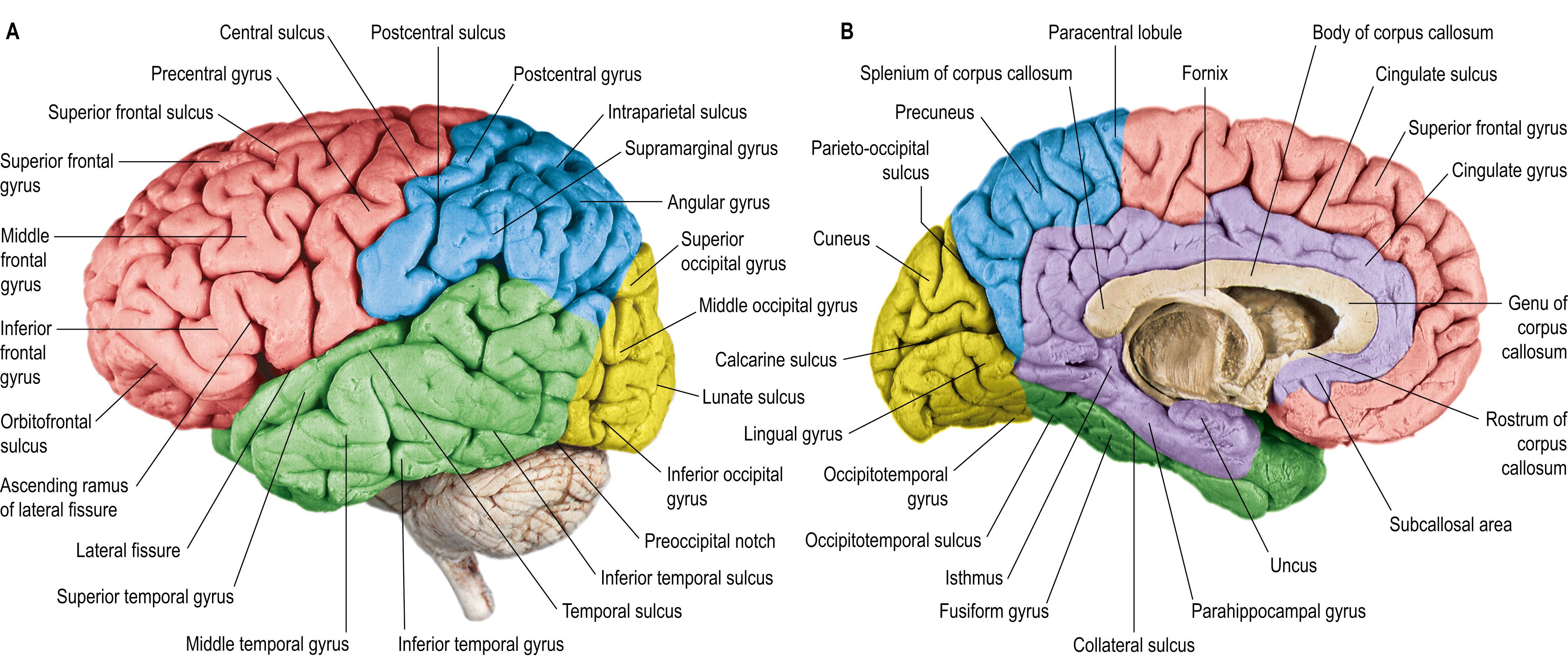
cerebral cortex gyri and sulci
The brain’s wrinkles are not arbitrary decoration—they are the visible outcome of development, mechanics, and evolutionary design working together.
THE WRINKLE MYSTERY: WHAT WE'VE LONG WONDERED
Humans and many other mammals do not have smooth brains. From dolphins and elephants to primates, the cerebral cortex—the outer layer that supports perception, language, and abstract thought—folds so that a large cortical surface can fit inside a limited skull volume. That much has been known for a long time. But pinning down the precise causes of folding has been contentious.
Two broad classes of explanations dominated scientific thinking: genetic and mechanical. Genetic models emphasize molecular cues and cellular programs that pattern the cortex during development. Mechanical models, by contrast, treat folding as an emergent property—an outcome of differential growth rates, physical forces, and simple buckling when one layer expands faster than another. Each view provided important pieces of the puzzle, but neither alone explained everything researchers observed across species, developmental stages, and pathological conditions.
HOW FOLDS FORM: THE MECHANICS OF GYRIFICATION
Physical forces and buckling
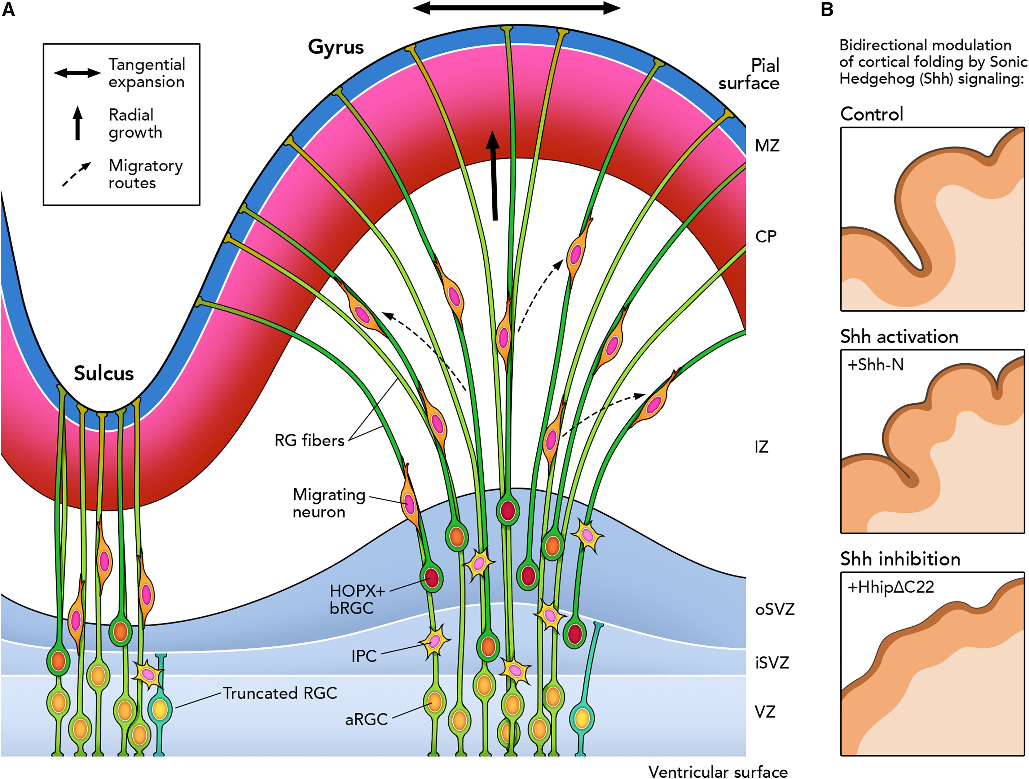
Imagine trying to fit a large sheet of paper into a small box: the excess surface must crumple, crease, or fold. The developing cortex behaves similarly. As neural progenitor cells multiply and generate neurons, the cortical sheet expands tangentially. If the layer beneath (the subcortex) does not expand at the same rate or resists movement, the cortical layer will buckle into folds.
This mechanical view casts gyrification as a physics problem. Regions that grow more rapidly—because of local cell proliferation or migration—become outward bulges (gyri). Adjacent areas that expand less become sulci. Mathematical models and soft-tissue experiments have shown that differential growth can reproduce many folding patterns seen in brains, including the characteristic spacing and curvature of gyri and sulci.
Tension along axons: wiring pulls the map
Another mechanical idea is axonal tension. As neurons send long-range connections across the cortex, the bundled axons exert pulling forces that could fold the tissue into patterns that reduce wiring length and energy cost. This hypothesis helps explain why functionally related areas lie near each other, and why folding tends to respect functional boundaries. Though elegant, measurements of tension in living tissue and computational studies indicate that axonal pulling alone is unlikely to account for the full complexity of cortical folding.
GENES AND CELLS: THE MOLECULAR DRIVERS
Progenitors, basal radial glia, and regional expansion
Genetic factors matter. During fetal development, the behavior of neural progenitor cells—how many divide, how long they proliferate, and where their progeny migrate—directly shapes the size and thickness of cortical regions. A specialized class of progenitors known as basal radial glia (bRG) is especially important in species with highly folded brains. These cells amplify the number of neurons and drive expansion of the cortical surface.
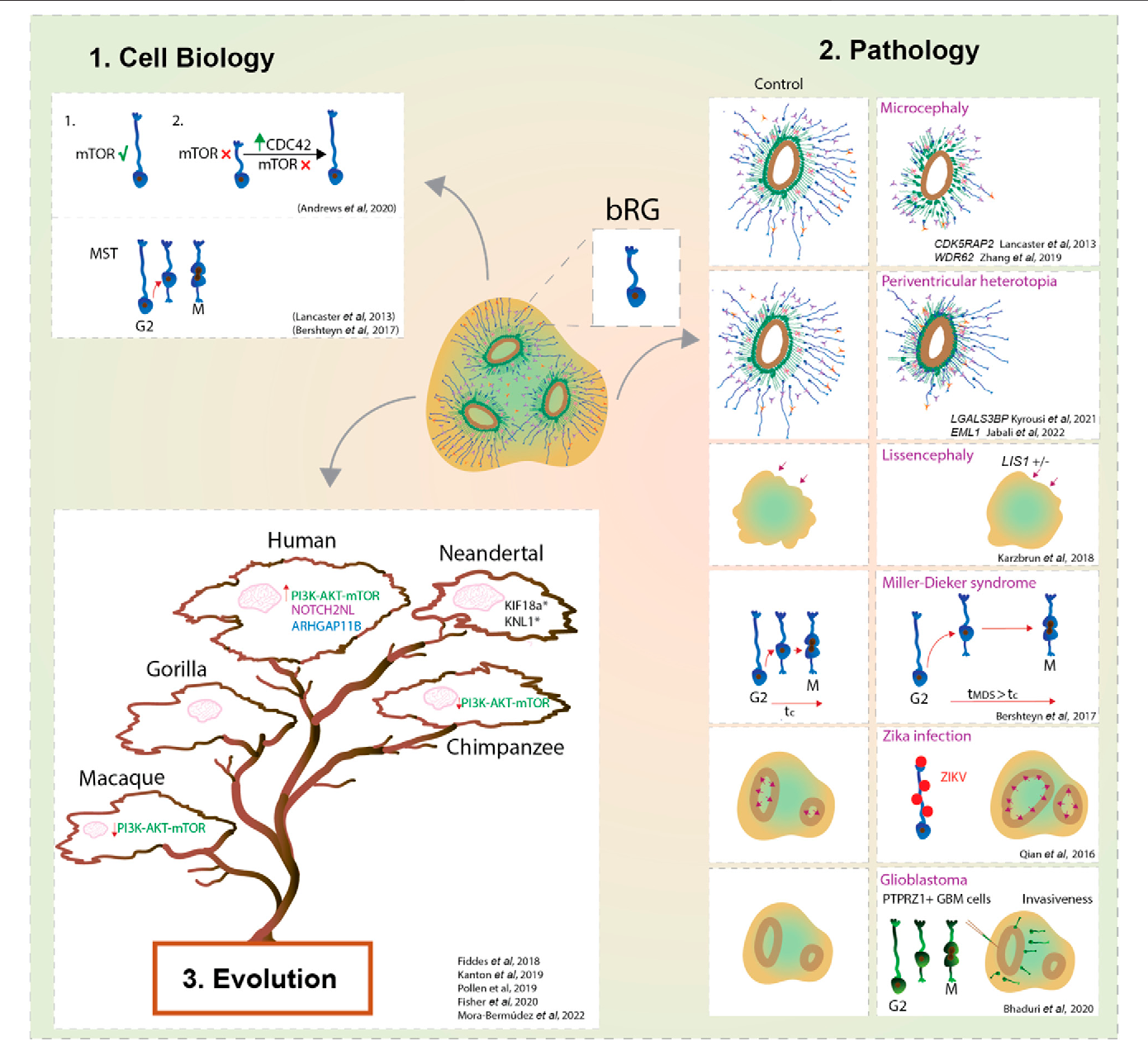
basal radial glia cells
Experimental manipulation of genes that control progenitor proliferation produces dramatic changes in folding. Increasing the proliferative capacity of the cortex in model animals can induce gyrification in otherwise smooth-brained species; conversely, mutations that reduce progenitor proliferation can yield smoother brains and serious neurological disorders.
Molecular switches: how a single protein can alter folding
Researchers have identified molecular regulators—transcription factors, growth factors, and cytoskeletal proteins—that bias development toward greater tangential expansion or thicker cortex. Small changes in expression levels or timing can have outsized effects on how and where folds appear. These molecular cues interact with mechanical forces: genes set the stage, and physics performs the choreography.
A SURPRISING NEW MECHANISM
The discovery in plain language
What made the recent discovery notable was its hybrid character: scientists found a mechanism that links cellular behavior to emergent mechanical forces in a way that was previously underappreciated. Without naming specific studies here, the core finding was that layers of cells within the developing cortex can undergo localized stiffness changes and reorganize their internal architecture, creating microdomains that bias where buckling initiates. In other words, cells don’t just grow faster or slower across broad regions—they can locally change the tissue’s mechanical properties to steer the folding process.
This mechanism explains why certain sulci are reliably positioned across individuals and species even when overall growth rates vary. It also clarifies how small, genetically-encoded changes in cell behavior produce reproducible macroscopic patterns of folds.
How this ties genetics to mechanics
By altering cytoskeletal tension, extracellular matrix composition, or cell-cell adhesion in targeted microdomains, genes can create predictable mechanical heterogeneities. Those heterogeneities act as 'weak spots' or 'seeds' for buckling when global growth reaches a threshold. The result is a folding pattern that is both genetically guided and physically emergent.
Small cellular shifts—changes in stiffness, adhesion, or extracellular matrix—can steer the brain's large-scale folding patterns.
WHY FOLDS MATTER: FUNCTIONAL AND EVOLUTIONARY SIGNIFICANCE
Surface area, connectivity, and computation
The simplest advantage of folding is geometric: folds increase cortical surface area without requiring a proportionally larger skull. More surface area allows for more neurons and synaptic connections, supporting higher computational capacity. But folds aren’t just passive folding of extra tissue; their arrangement influences connectivity. Neighboring gyri often contain related functional maps, and sulcal boundaries frequently align with transitions in cytoarchitecture and function.
Evolutionary comparisons show that gyrification correlates imperfectly with intelligence but robustly with cortical expansion and certain ecological demands. Some species evolved highly folded cortices alongside specialized sensory or social skills. Folding patterns thus reflect a mixture of developmental constraints, genetic programs, and adaptive pressures.
Clinical relevance: why surgeons and neurologists care
For clinicians, understanding folding is essential. Many neurodevelopmental disorders feature abnormal gyrification. Lissencephaly, literally “smooth brain,” results from severe failure of folding and is associated with profound developmental delay and epilepsy. Other conditions, like polymicrogyria, microgyria, or focal cortical dysplasia, involve aberrant folding or cortical organization and can underlie seizures and cognitive deficits.
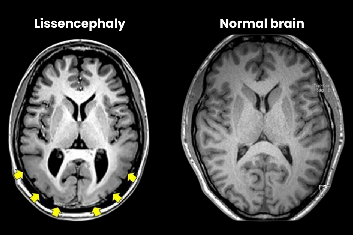
lissencephaly smooth brain MRI
Better knowledge of folding mechanisms can improve prenatal diagnosis, inform surgical planning, and suggest therapeutic targets to prevent or mitigate developmental disorders.
WHEN FOLDING GOES WRONG
Lissencephaly, polymicrogyria, and other disorders
Genetic mutations that impair neuronal migration, progenitor proliferation, or structural proteins can prevent normal folding. Lissencephaly produces a markedly smooth cortex and frequently arises from mutations in genes that guide neuronal migration. Polymicrogyria, characterized by excessively many small folds, often reflects insults occurring later in gestation, such as ischemia or infection, and can produce focal neurological symptoms.
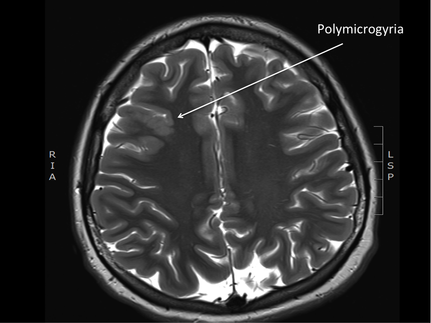
polymicrogyria brain pathology
Understanding the newly discovered microdomain mechanism gives researchers a fresh lens to interpret these disorders: altered stiffness or adhesion in specific cortical patches could seed pathological folding patterns. That perspective may open new strategies for early intervention.
IMPLICATIONS FOR RESEARCH AND MEDICINE
Model systems and translational opportunities
The discovery urges a rethinking of model systems. Brain organoids—miniature, simplified brain models grown from stem cells—have become a popular way to study human cortical development. If local mechanical heterogeneities play a major role, researchers must ensure organoids faithfully replicate not just cellular composition but tissue mechanics and matrix properties. That insight also suggests potential therapeutic strategies: modulating extracellular matrix composition or cell adhesion pharmacologically during critical windows might correct or reduce abnormal folding in high-risk fetuses.
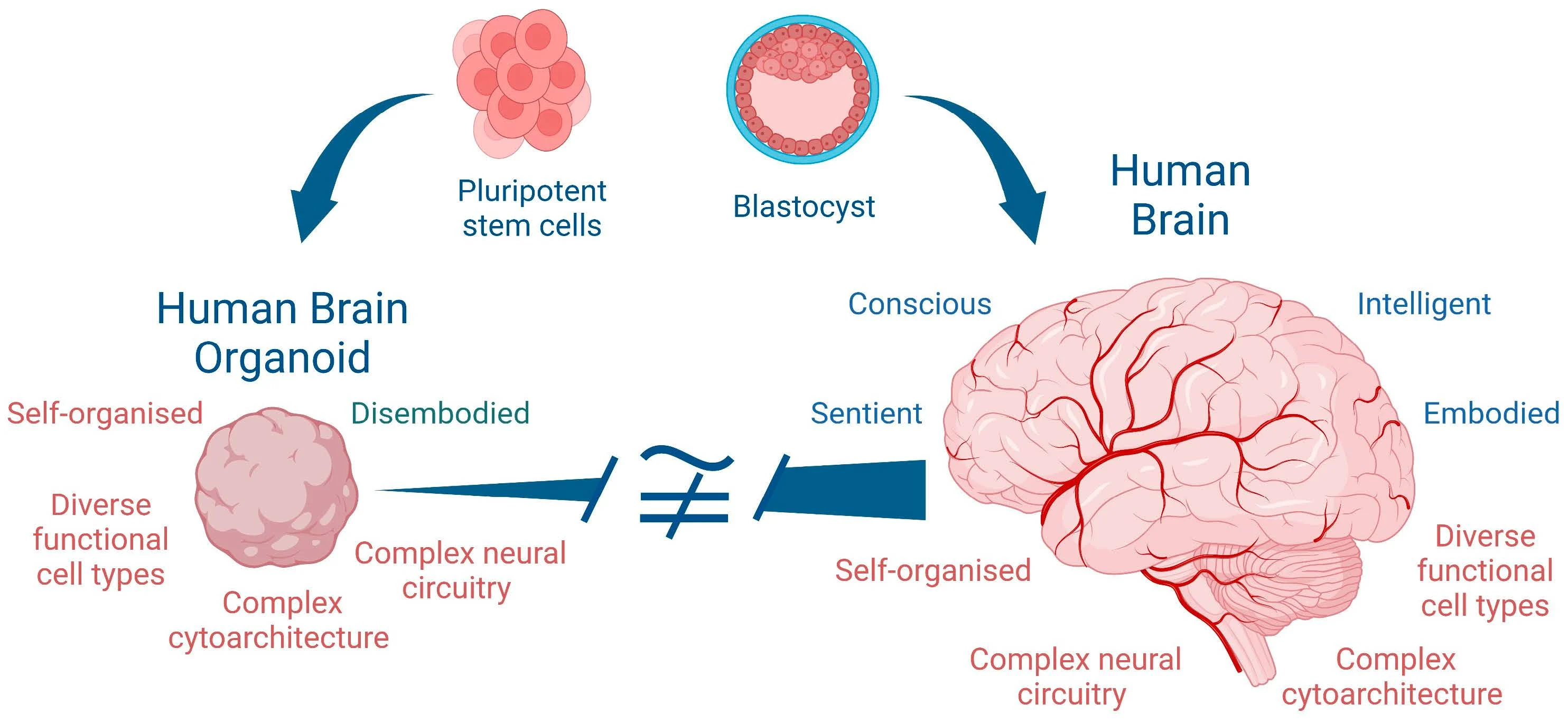
brain organoid stem cells
Connecting development to adult diversity
Folding patterns also vary between healthy individuals, and that variation may underlie subtle differences in cognition, personality, or vulnerability to disease. The interplay between genes and mechanics provides a principled way to study how small developmental tweaks can cascade into lifelong neuroanatomical diversity.
PRACTICAL TAKEAWAYS FOR THE CURIOUS READER
What the discovery changes in simple terms
- It connects the dots: Folding is not just physics or genetics, but a dialogue between the two mediated by cell-level mechanics.
- It refines models: Predictive models must include local tissue stiffness and adhesion patterns as well as growth rates.
- It suggests interventions: Understanding tissue mechanics could open new prenatal or early-life therapies for cortical malformations.
The brain’s landscape is shaped by a conversation between genes and the laws of physics.
LOOKING AHEAD: OPEN QUESTIONS
As with any discovery, the new mechanism raises more questions than it answers. How exactly are localized stiffness patterns encoded genetically? What molecular pathways control the extracellular matrix and cell adhesion with the required spatial precision? How plastic are folding decisions—can interventions in late gestation or infancy alter patterns in predictable ways? And how do these microdomain-driven folds interact with axonal tension and large-scale growth dynamics?
Answering these questions will require interdisciplinary work spanning developmental biology, biomechanics, computational modeling, and clinical research. It will also demand careful ethical consideration when moving toward prenatal interventions.
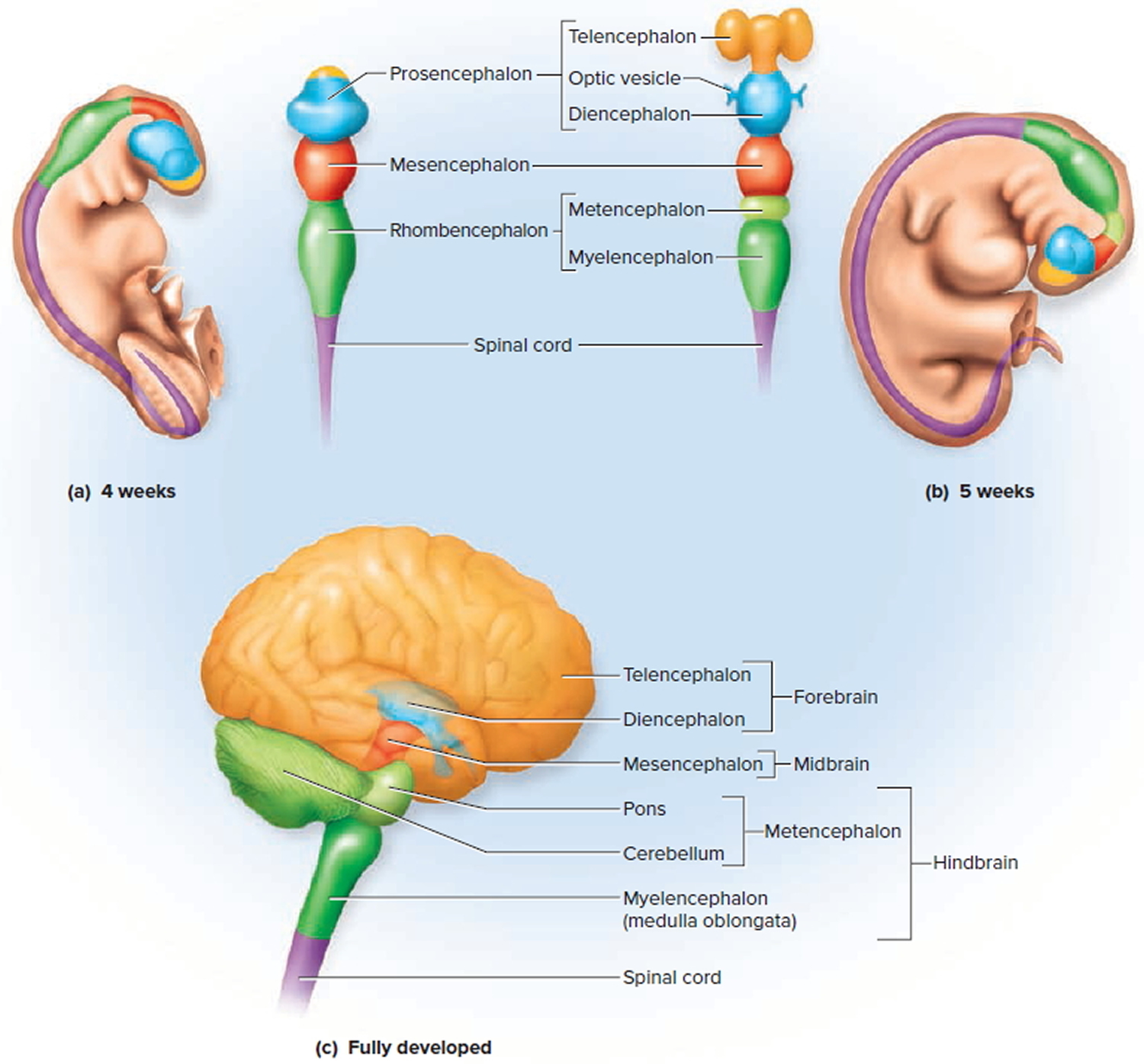
fetal brain development stages
CONCLUSION: WHY THIS MATTERS
The newly identified mechanism does not overthrow prior ideas; it enriches them. Folding arises from a multilayered process in which genes specify cellular behaviors that create mechanical heterogeneities, and those heterogeneities guide where the cortex buckles as it grows. That synthesis helps explain features of both normal variation and pathological malformation, and it points to new directions for research and clinical care.
At a deeper level, the story of the brain’s wrinkles is a reminder of how biological form emerges: through the interplay of information encoded in DNA, the lived dynamics of cells and tissues, and the immutable laws of physics. The next time you gaze at a brain image, you can appreciate those grooves and ridges not only as anatomy, but as a palimpsest of development, evolution, and the forces that shaped the organ that makes thought possible.
- Brain folding (gyrification) results from both genetic programs and mechanical forces; recent work highlights local tissue mechanics as a decisive factor.
- Localized changes in stiffness, adhesion, or matrix composition can seed predictable folds when the cortex expands.
- Understanding folding helps explain neurodevelopmental disorders and suggests new avenues for diagnosis and therapy.
Illustrations and simplified diagrams can help readers visualize folding mechanics—look for fetal brain atlases or interactive models for a clearer view.