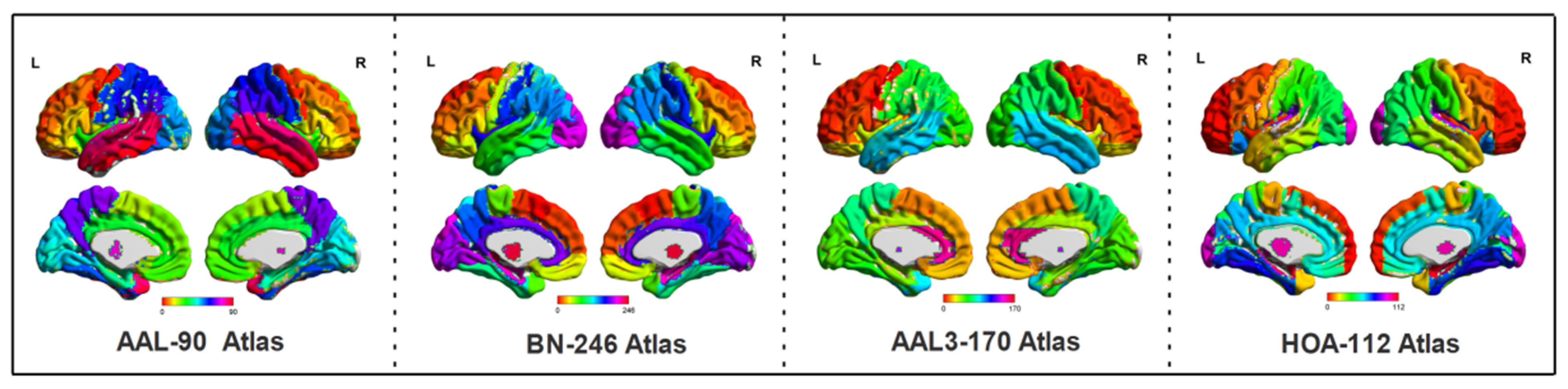
A Hidden Map of the Human Brain: 539 Distinct Regions
The headline is elegant and deceptively simple: scientists report a brain map that divides the human brain into 539 distinct regions. What looks like a numerical milestone is in fact the culmination of decades of effort to tease apart how structure, connectivity and function combine to produce the mind. This new atlas—built from large-scale imaging, careful anatomy, and advanced computational modeling—promises to change how researchers design experiments, how clinicians plan surgeries, and how we think about the brain’s organizational logic.
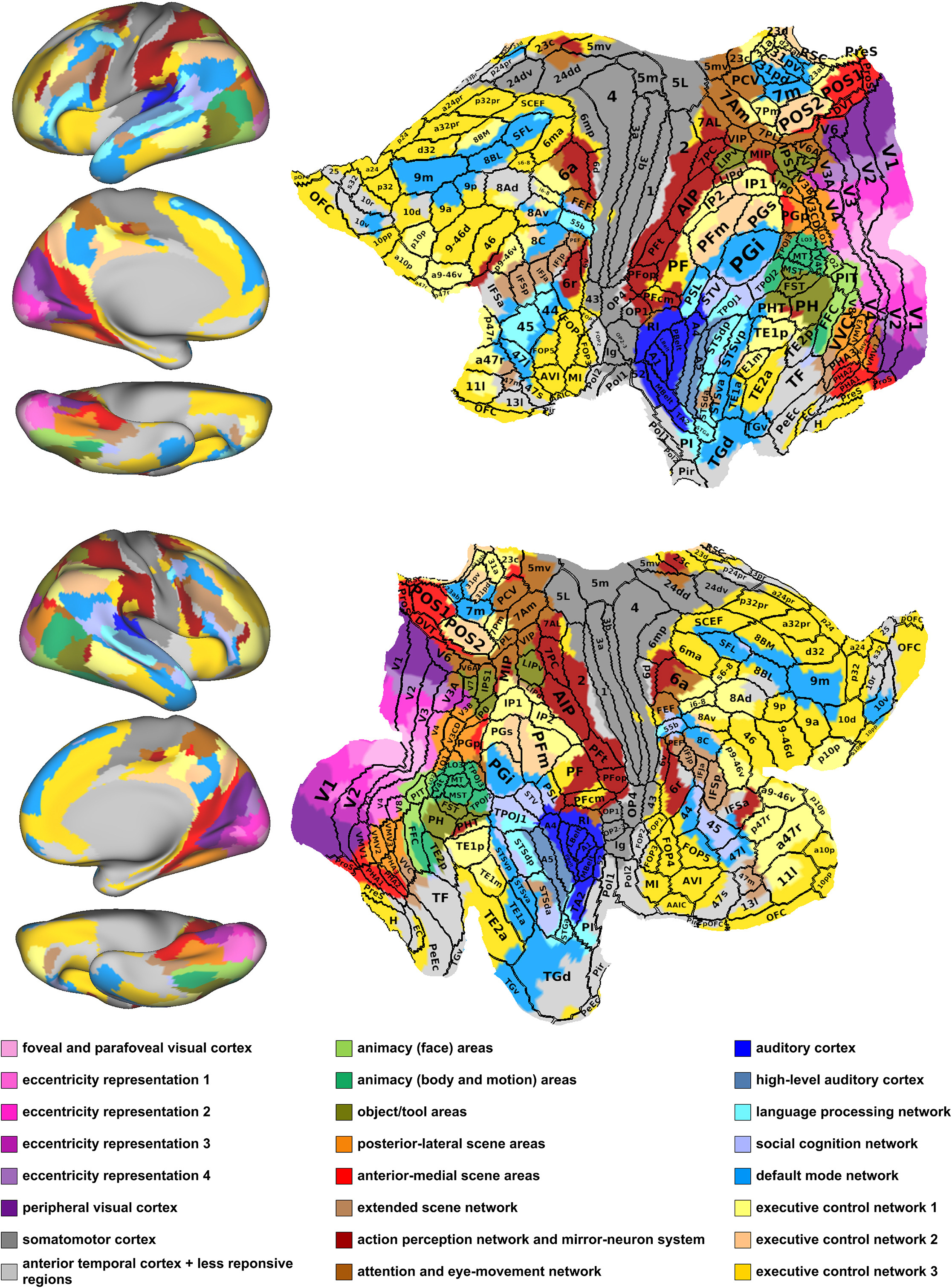
human brain cortical parcellation map
This atlas is less an endpoint than a new lens: it sharpens where we look and how precisely we can measure change across health, behavior, and disease.
WHY A NEW ATLAS MATTERS
The brain is not a homogeneous blob. For more than a century neuroscientists have tried to parcel it into meaningful units—areas that share cellular architecture, patterns of connectivity, or consistent functional responses. Early maps, like Brodmann’s areas, used postmortem anatomy to define roughly 50 regions. More recently, large-scale projects using magnetic resonance imaging identified hundreds of cortical parcels—such as the Human Connectome Project’s 360-region parcellation. A 539-region atlas expands that precision and integrates both cortical and subcortical detail in ways that are immediately useful.
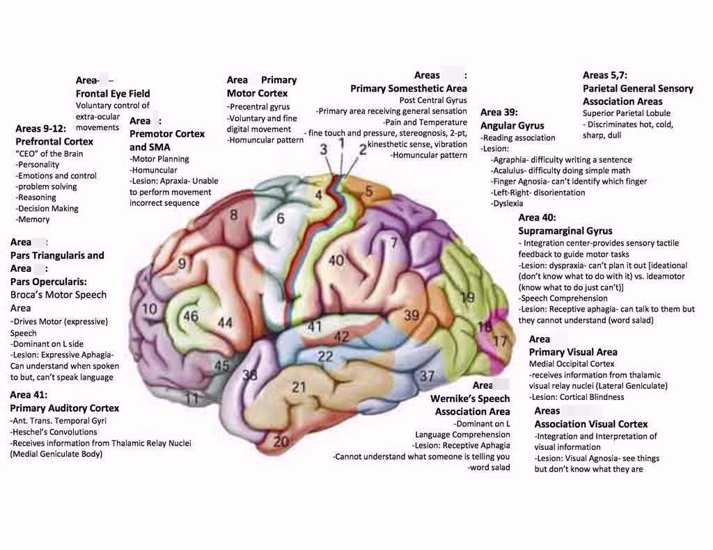
Brodmann areas brain map diagram
Why should anyone outside a lab care? Because how you define a ‘region’ drives every measurement that follows. Biomarkers, surgical targets, neural stimulation sites, and machine-learning models all depend on the units of analysis. A finer-grained, biologically informed atlas reduces ambiguity and makes comparisons across studies far more reliable.
HOW THE MAP WAS BUILT
Multimodal imaging: marrying structure and function
Creating a map is not simply dividing the brain into equally sized tiles. The most convincing parcellations rely on multiple sources of information—structural anatomy (how cells and layers are organized), functional MRI (how activity patterns correlate during tasks and rest), and diffusion MRI (how white-matter tracts physically connect regions). Integrating these modalities helps ensure that parcels are homogeneous across many biological dimensions rather than just one noisy measurement.
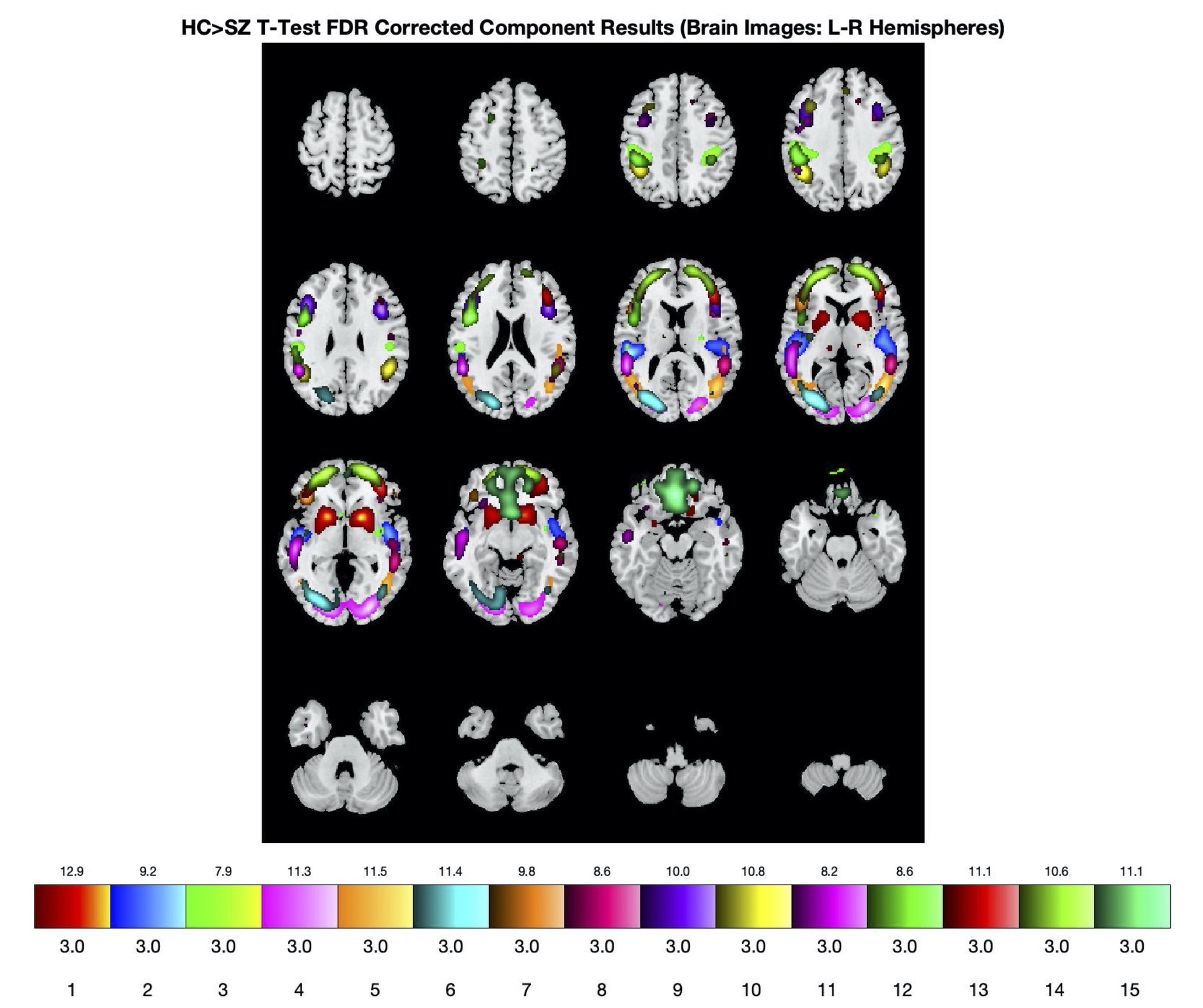
fMRI functional brain connectivity scan
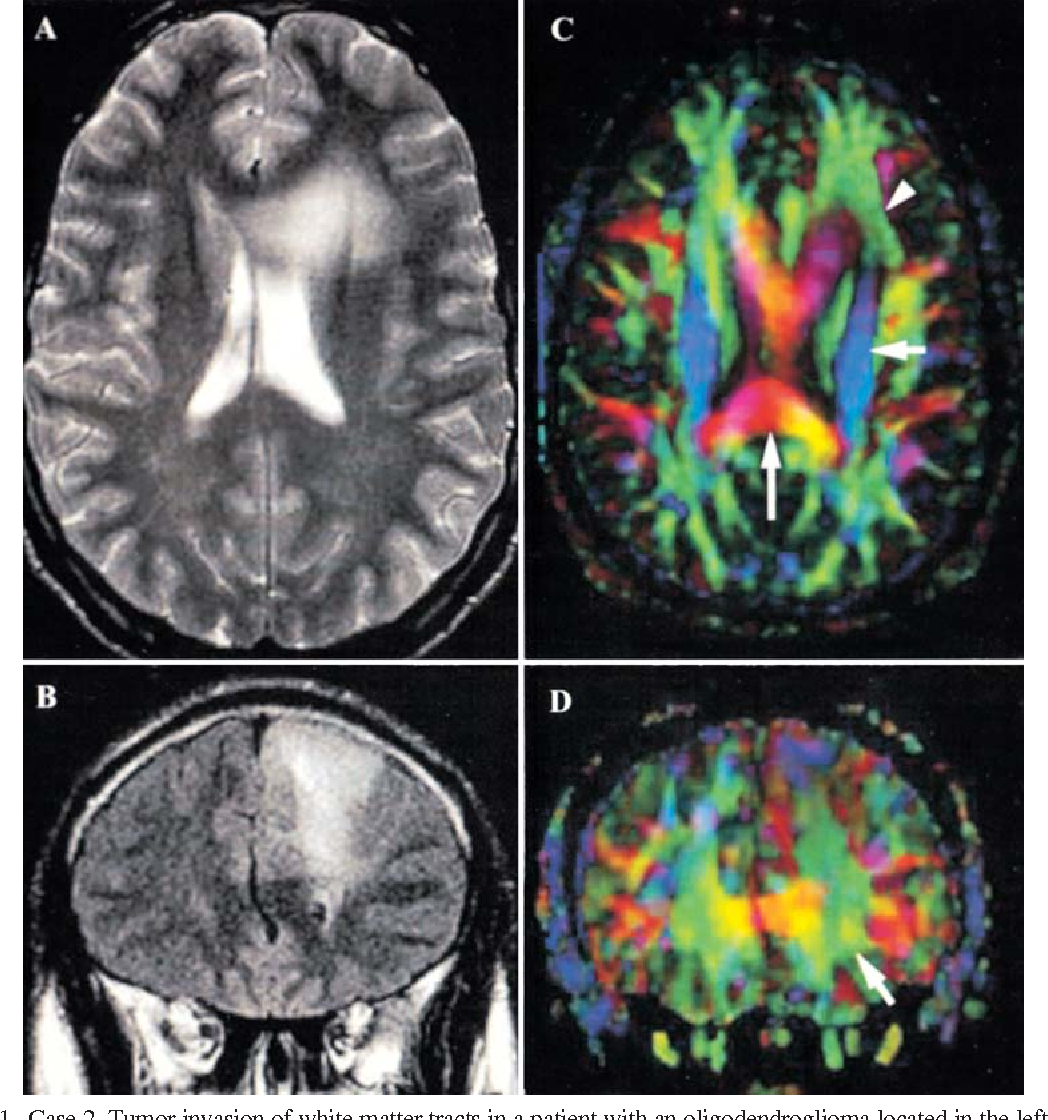
diffusion MRI white matter tracts
Advanced analytics and reproducibility
Computational methods—clustering, gradient analysis, and machine learning—are applied to multimodal datasets to find boundaries where properties change sharply. Importantly, the best maps are evaluated for reproducibility: do the same parcels appear when you analyze different groups of people, different scanners, or slightly different preprocessing choices? A robust atlas includes statistical measures of reliability and confidence for each parcel.
Large datasets and population variation
Modern atlases leverage thousands of high-quality scans to capture common structure while acknowledging individual differences. The 539-region map likely incorporates population-level templates and probabilistic labels, meaning each region comes with a sense of how consistently it appears across people, and where individual anatomy might deviate.
WHAT THE 539 REGIONS REPRESENT
Cortex, subcortex, and transitional zones
The new map almost certainly spans cortical areas (the outer folded sheet of neurons), subcortical nuclei (deep structures like the thalamus and basal ganglia), and cerebellar regions. It also highlights transitional zones—areas where functional properties blend or where structural boundaries are unclear. Including these transition regions is crucial; many cognitive functions arise at interfaces between canonical networks.
Functional specialization at higher resolution
Breaking a previously defined region into two or more smaller parcels might reveal that what was once thought to be a single functionally uniform area actually contains distinct subunits with separate connectivity profiles and task responses. For instance, a visual area might split into subregions with preferences for central vs peripheral vision, or a prefrontal parcel might separate cognitive-control and social-evaluation subunits.
IMPACT ON RESEARCH AND MEDICINE
Sharper biomarkers for psychiatric and neurological disorders
Many studies seek brain-based biomarkers for conditions such as depression, schizophrenia, Alzheimer’s disease, and epilepsy. Coarser parcellations can blur disease signals; smaller, well-defined regions can reveal subtle differences in connectivity or activity that better predict diagnosis, prognosis, or treatment response. Clinicians and researchers can use this level of detail to stratify patients and tailor interventions more precisely.
Improved neurosurgical planning and stimulation targeting
Surgery and neuromodulation rely on precise anatomy. For tumor resection, preserving essential functional tissue depends on accurate maps of language, motor, and sensory areas. Deep brain stimulation (DBS) and noninvasive stimulation techniques (like TMS and tES) require sub-centimeter precision to modulate targeted circuits. A 539-region atlas gives clinicians more refined coordinates and probabilistic guidance about where function actually resides.
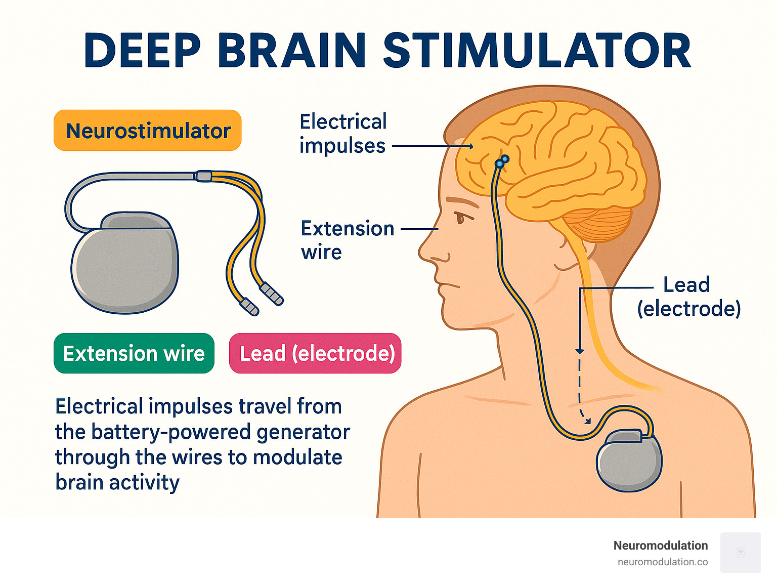
deep brain stimulation electrode anatomy
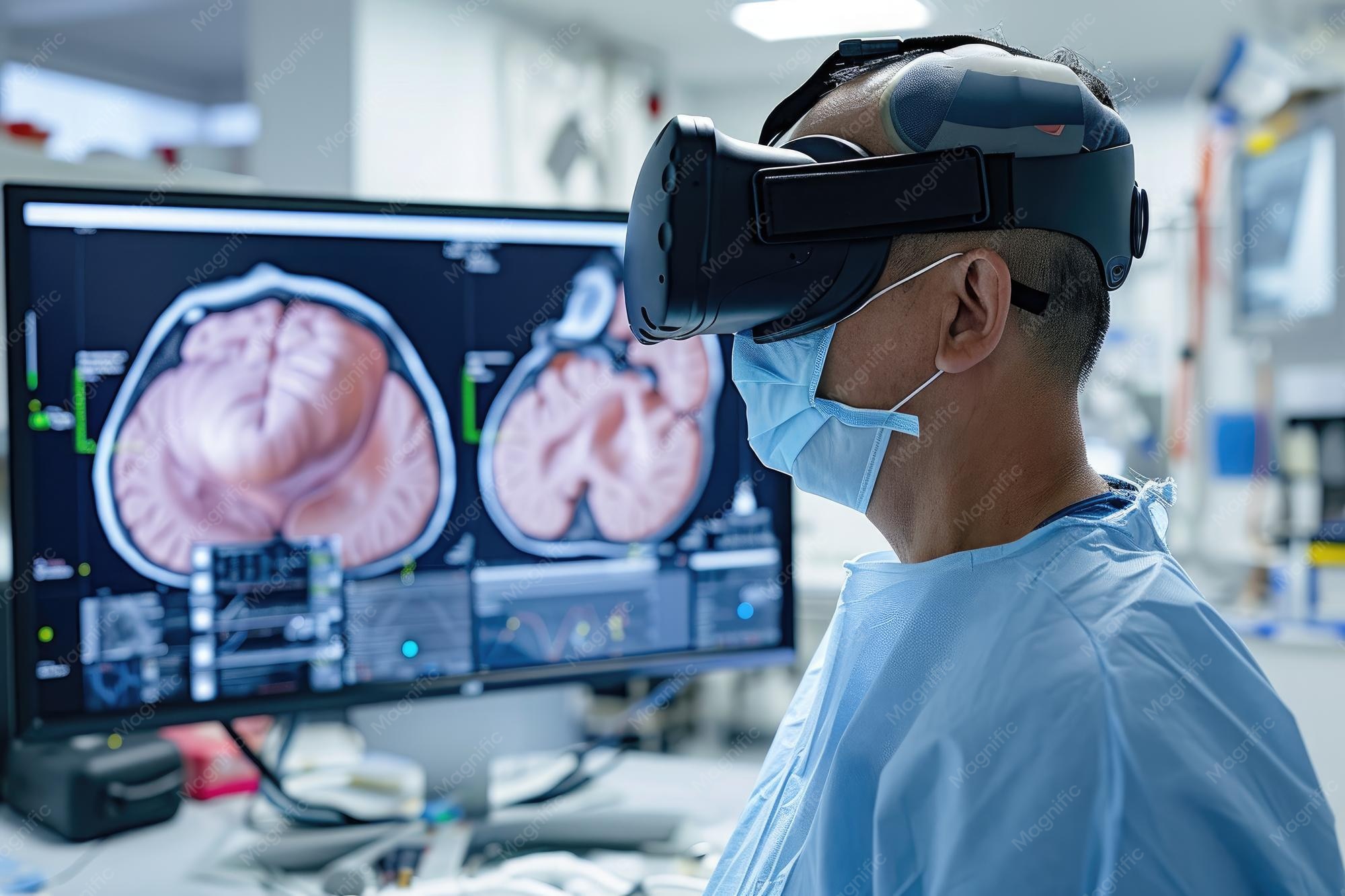
neurosurgeon planning brain surgery screen
Better models of cognition and brain networks
Computational models of cognition—from network simulations to machine-learning classifiers—benefit when input nodes correspond to biologically meaningful units. With more precise nodes, models can capture richer dynamics, such as how local microcircuits interact with distributed networks to produce behavior. That, in turn, refines theories about memory, attention, language, and perception.
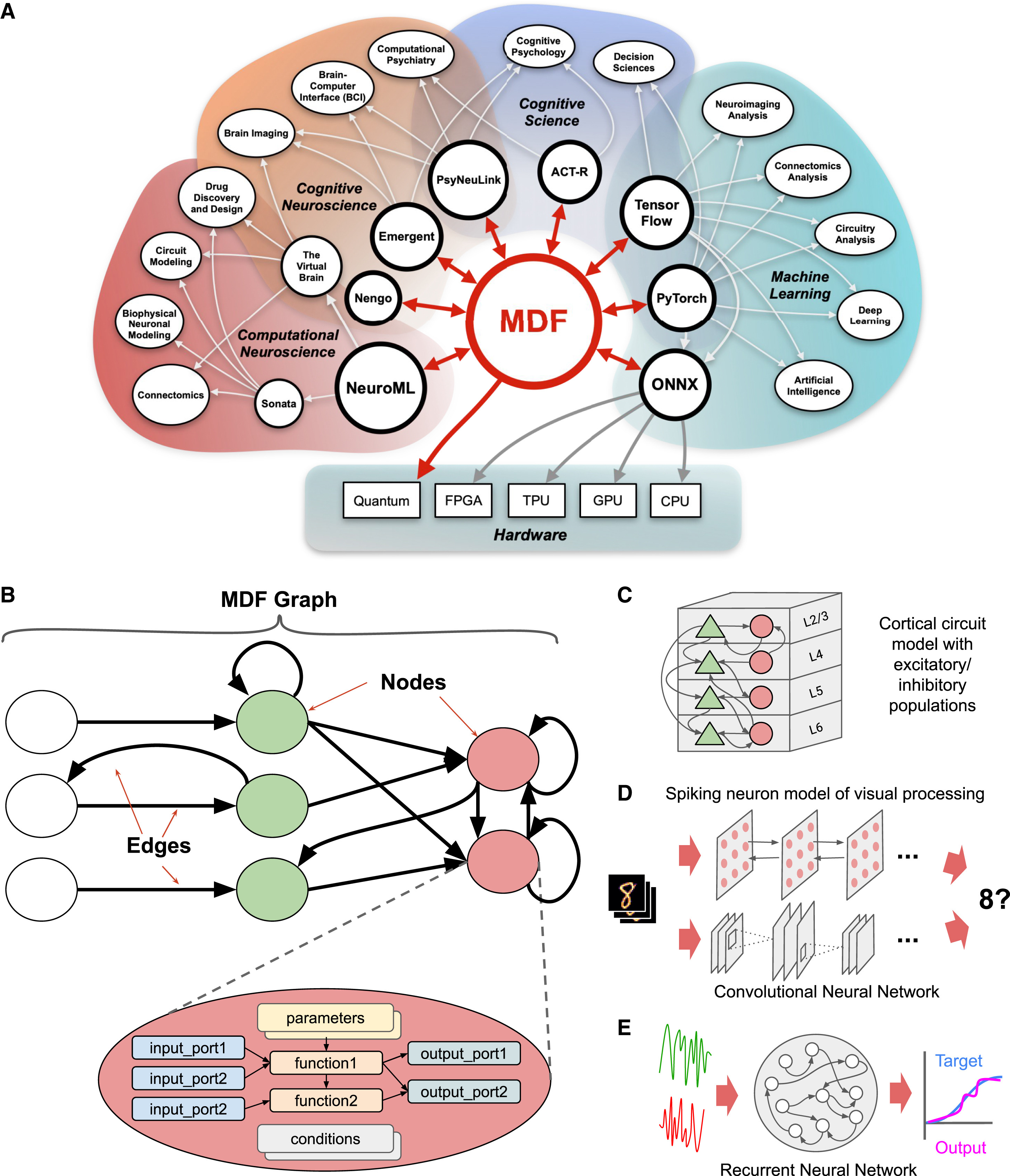
computational neuroscience brain network model
ARGUMENTS AND CAUTIONS
Is more always better?
Granularity is a trade-off. Very fine parcellations can overfit noise or capture idiosyncratic variations that are not biologically meaningful. Researchers must ask: does splitting an area add explanatory power for the question at hand? If not, finer granularity can complicate analysis and interpretation.
Reproducibility across scanners and populations
Imaging protocols, preprocessing pipelines, and population demographics can all affect parcellation. A parcel that is clear in one dataset might be ambiguous in another. High-quality atlases therefore report measures of cross-site reproducibility and provide guidance about appropriate use.
HOW THIS FITS INTO THE LEGACY OF BRAIN MAPS
Maps have guided neuroscience since Brodmann, who used cytoarchitecture to delineate roughly 50 areas. The 20th and 21st centuries expanded that view: functional imaging identified distributed networks; diffusion imaging charted axonal highways; and big-data projects created high-resolution cortical parcellations. The 539-region atlas is the next layer, integrating modalities and offering probabilistic labels that reflect population variability.
A brief comparison
Previous landmark parcellations varied in scope and intent—some prioritized cytoarchitecture, others functional homogeneity, and still others connectivity. The current move toward multimodal parcellations reflects the understanding that structure, function, and connectivity are complementary. Where earlier atlases gave us broad provinces, newer atlases aim for neighborhoods and city blocks.
PRACTICAL ADVICE FOR RESEARCHERS
Choosing the right parcellation
Researchers should choose an atlas based on the question: coarse parcellations may suffice for whole-brain network topology; finer maps are better for localizing function or detecting subtle disease changes. It is often valuable to run sensitivity analyses across multiple atlases to ensure findings are robust.
Integrating individualized mapping
Group atlases are helpful, but individual brains vary. Ideally, studies will combine a population-derived atlas with individualized registration or functional localizers that refine parcel boundaries in each subject.
THE FUTURE: WHERE NEXT?
Towards multilayer atlases
The future likely holds atlases that layer multiple scales simultaneously—from microstructural gradients and cell-type maps to mesoscale regions and macroscale networks. Such multilayer atlases will help link genes and molecules to circuits and cognition.
Clinical translation and predictive tools
As atlases mature, they will be integrated into clinical workflows—helping predict who will respond to a drug, where to target stimulation, or when a surgical approach risks critical function. Machine-learning models trained on atlas-informed features could power decision-support tools in the clinic.
POTENTIAL LIMITATIONS AND OPEN QUESTIONS
Even a detailed atlas leaves many questions unanswered. How stable are parcels across the lifespan? Do developmental factors or neuroplastic changes substantially reshape boundaries? How do individual differences in microanatomy map onto atlas parcels? And crucially, how should researchers interpret negative findings when a hypothesized effect is absent at one level of parcellation but present at another?
CONCLUSION
The report of a 539-region brain atlas is a landmark not because it is the final map but because it amplifies our capacity to measure and ask better questions. It bridges modalities, quantifies uncertainty, and provides a practical scaffold for both basic science and clinical translation. As with any map, its value depends on how wisely it is used: as a guide, not a decree.
- Precision matters: A 539-region atlas offers finer-grained, probabilistic parcels that can improve biomarker discovery and surgical targeting.
- Multimodal integration: The atlas combines anatomical, functional, and connectivity data to define regions that are biologically coherent.
- Trade-offs exist: Higher granularity improves localization but may increase sensitivity to noise and individual variability.
- Practical use: Researchers should match atlas granularity to their question and consider individualized mapping for clinical decisions.
The new atlas reframes questions about brain organization and sets the stage for a decade of more precise neuroscience.