Hidden Gut Defects Behind Recurrent IBD: Why It Keeps Returning
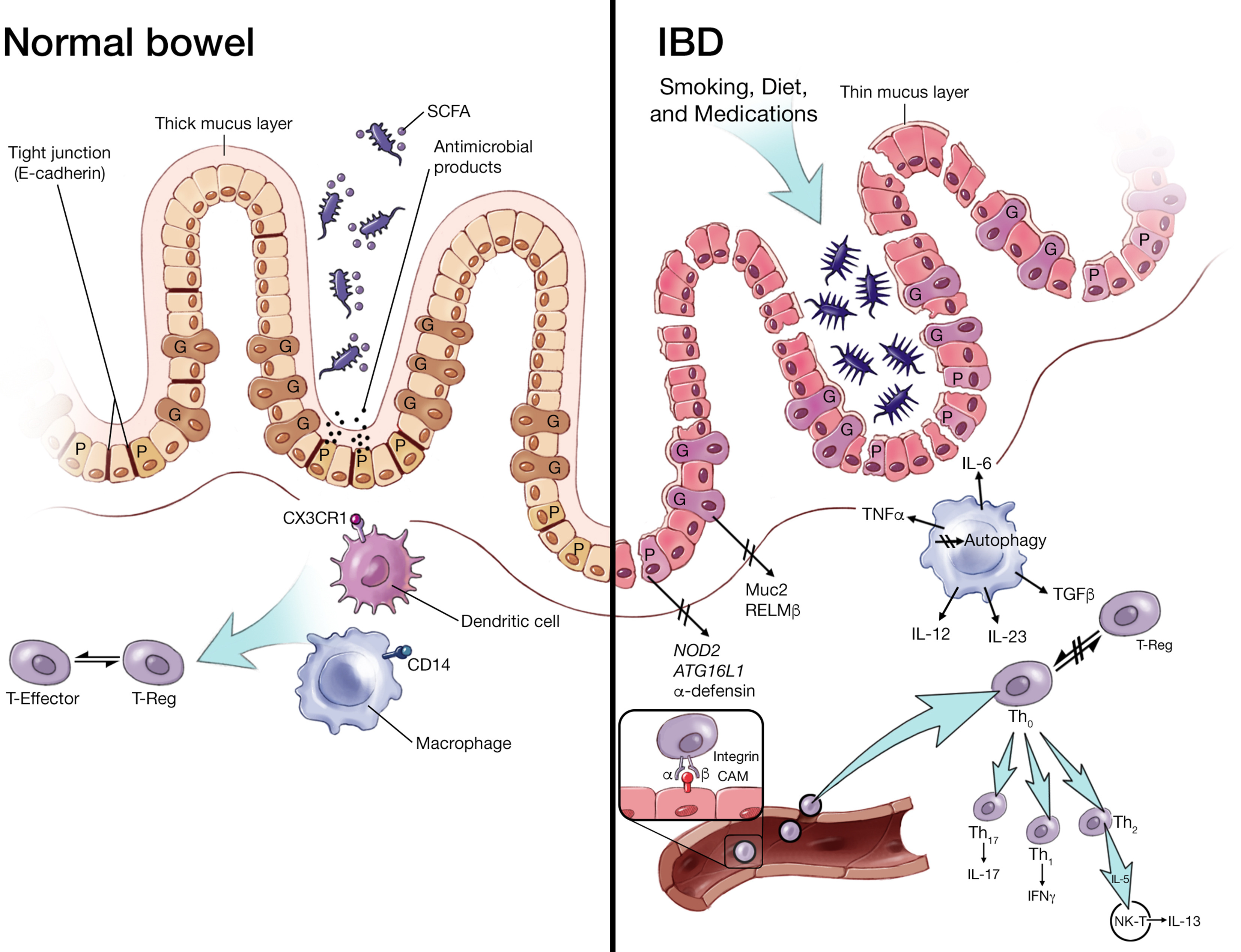
The familiar arc of inflammatory bowel disease—periods of quiet followed by a sudden flare—leaves patients and doctors asking the same blunt question: why does it come back? For many, the answer lies not in mood or diet alone, but in small, structurally or biologically concealed problems inside the bowel. These are not dramatic tumors or textbook fistulas; they are subtle defects and reservoirs that evade routine scans and superficial healing checks but seed inflammation months or years later. Understanding these hidden contributors changes how we diagnose, treat, and—crucially—prevent relapse.
How IBD Goes into Remission and Then Returns
IBD—an umbrella term for Crohn's disease and ulcerative colitis—behaves like a chronic wound that oscillates between control and flare. A combination of immune imbalance, genetic predisposition, microbial triggers, and environmental inputs sets the stage. Modern therapies, particularly biologic agents, are highly effective at damping immune activity and inducing clinical remission. Yet clinical remission and endoscopic remission are not always the same as durable healing of the entire intestinal wall or elimination of disease reservoirs. The difference is where these hidden defects matter.
Remission versus true tissue recovery
Even when symptoms subside and inflammation markers fall, pockets of microscopic inflammation, unhealed micro-ulcers, or biofilm-coated niches can persist. These micro-habitats harbor bacteria and inflammatory signals that can reignite symptoms when therapy is tapered or the immune system is challenged. In short, quiet does not always mean cured.
The Hidden Structural Defects That Often Get Missed
Clinicians are increasingly recognizing several recurring anatomic and mucosal problems that lie at the root of relapse. These include:
- Residual diseased bowel after surgery: incomplete removal of affected segments or diseased tissue at anastomosis sites.
- Small strictures and blind loops: narrowed areas that trap fecal matter and bacteria, creating a local nidus of inflammation.
- Micro-fistulas and sinus tracts: tiny channels that start deep in the bowel wall and escape detection on basic imaging.
- Biofilm reservoirs: protective bacterial communities that cling to micro-erosions and resist antibiotics and immune clearance.
- Persistent mucosal breaches: incomplete restoration of tight junctions and barrier proteins, leading to chronic permeability.
Individually or together, these defects act like embers under ash—hidden but very much capable of reigniting a full flare.
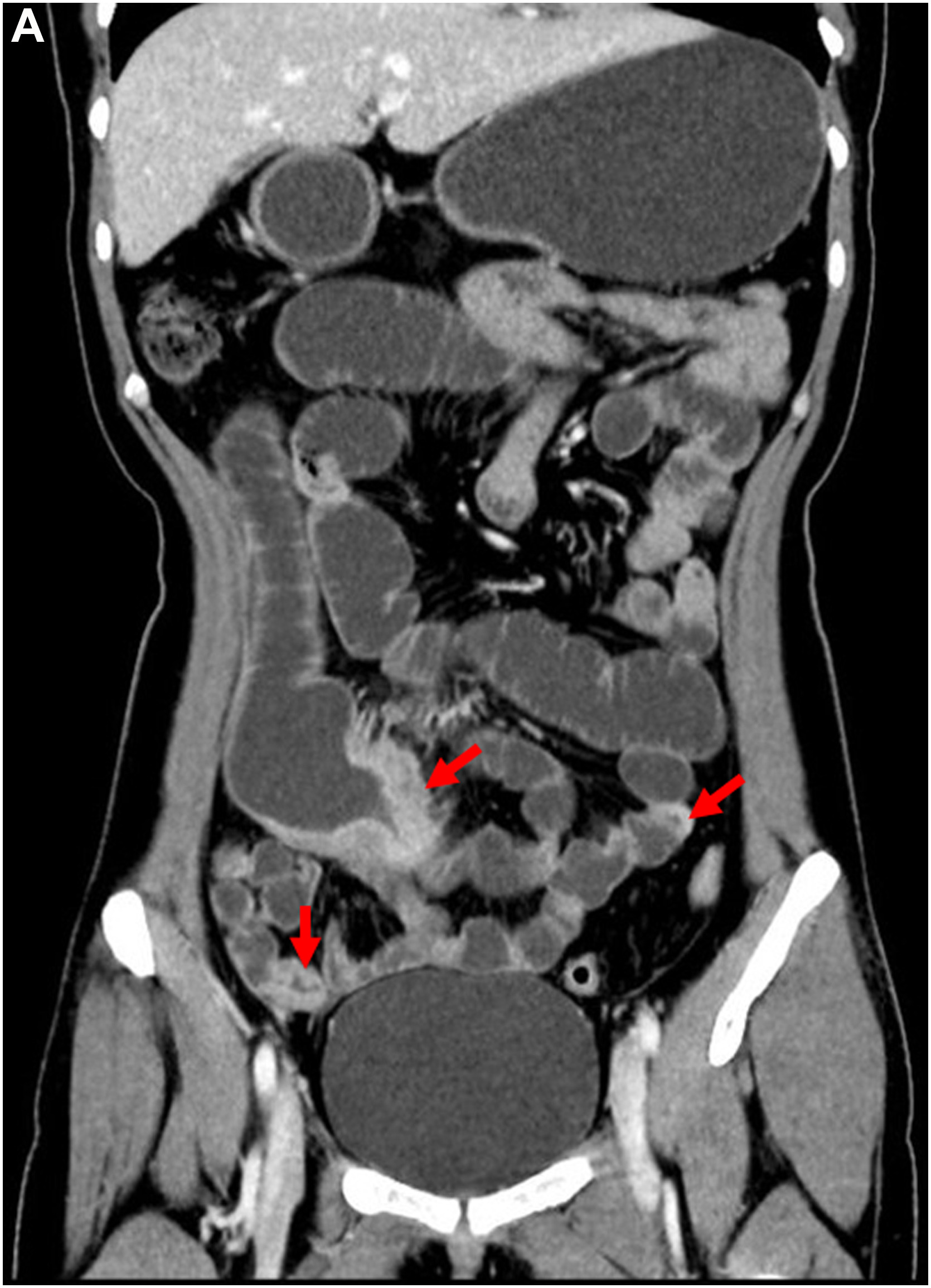
small bowel stricture IBD
Why routine tests sometimes fail to find them
Standard blood tests (CRP), symptom scores, and even conventional colonoscopy can miss these problems. CRP is nonspecific and can be normal in isolated small-bowel disease. Colonoscopy evaluates the mucosal surface, but deep transmural disease—common in Crohn’s—can lurk beyond the visible lining. Small, intermittent fistulas and biofilms aren’t always visible, and strictures that are functionally significant may be overlooked if they only cause subtle transit delay.
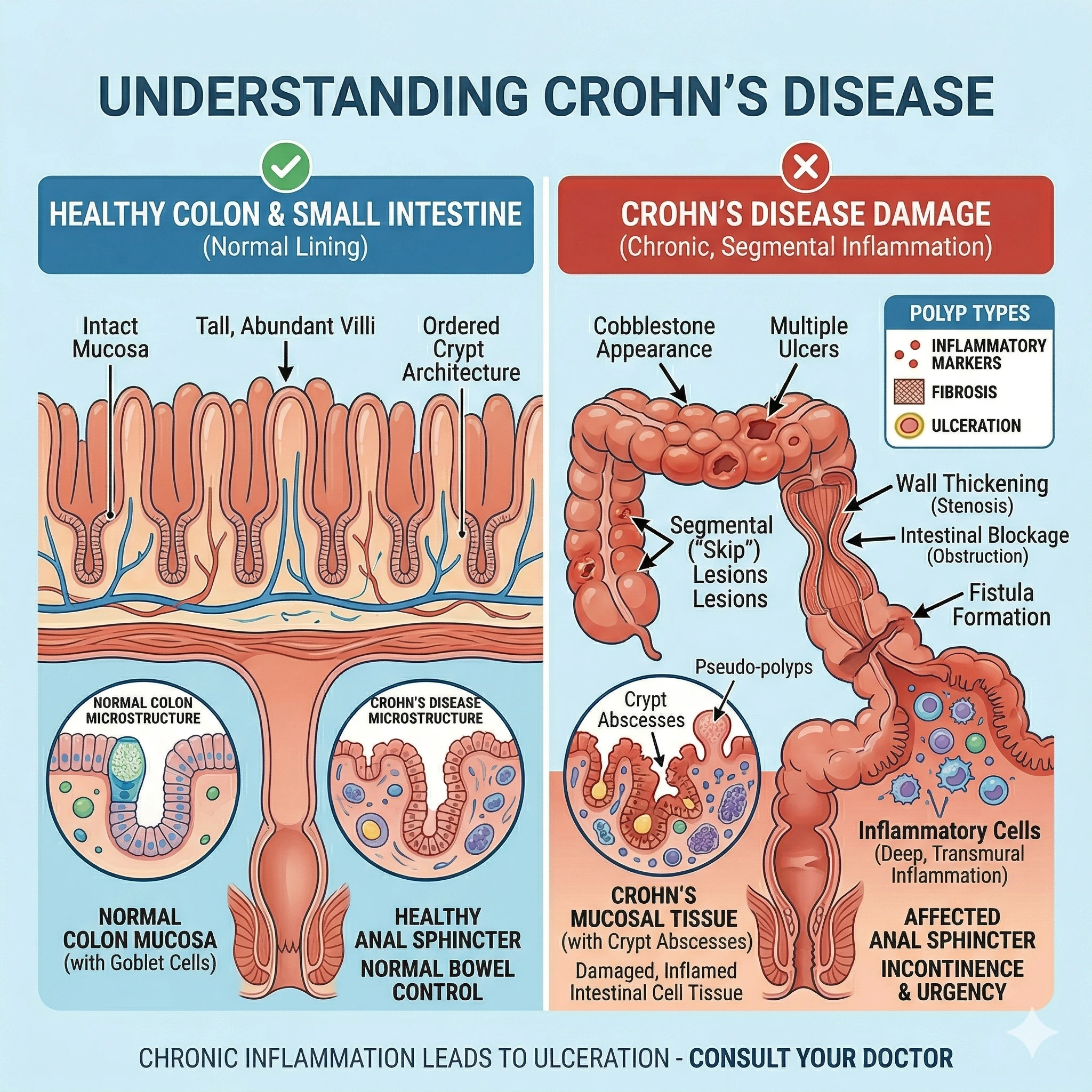
intestinal fistula Crohn
“You can achieve symptom control yet leave behind anatomic pockets that guarantee a return unless you look for them.”
Post-operative recurrence: an instructive model
Surgery illuminates the problem. After ileocolic resection for Crohn’s disease, recurrence at the anastomosis is common—often within a year—because microscopic disease persists at the margins or bacteria recolonize the surgical site. Understanding why recurrence happens here has broader lessons: even when major disease is removed, minor defects and altered anatomy create new environments that favor relapse.
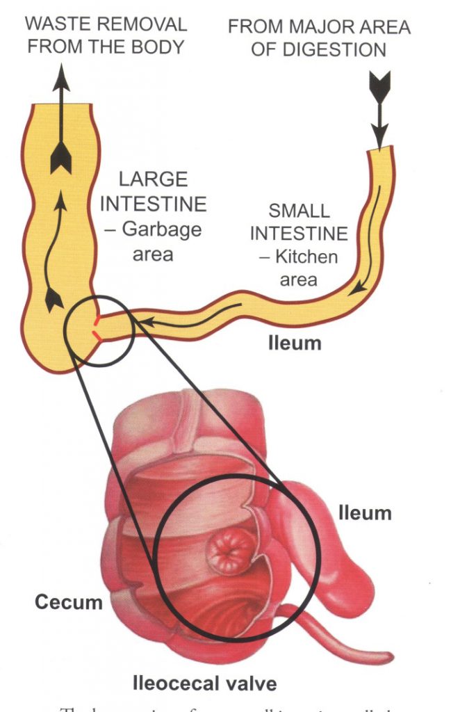
ileocecal valve Crohn disease
Anastomotic disease and biofilm
Surgical joins change local flow and mucosal exposure. Biofilms—sticky microbial communities—prefer these altered surfaces. They shield microbes from immune surveillance and from many antibiotics, becoming long-term irritants that promote inflammation. Recognizing anastomotic biofilm as a potential culprit changes follow-up strategy: look harder and earlier with targeted imaging and endoscopic surveillance.
The microbiome, barrier function, and immune memory
Inflammation isn't only structural; it’s biological. The gut barrier—composed of mucus, epithelial cells, and tight junctions—protects the immune system from microbial contact. When barrier proteins fail or mucosal healing is incomplete, translocation of microbes or their products stimulates a recurrent immune response. Meanwhile, the microbiome can reconfigure into a pro-inflammatory state that perpetuates disease even if the original trigger has been removed.
Biofilm and microbial persistence
Biofilms and dysbiotic communities can act as reservoirs of inflammatory triggers. They modulate local immunity, promote barrier dysfunction, and seed inflammation when systemic suppression is relaxed. Addressing these microbial reservoirs often requires a layered approach: targeted antibiotics, anti-biofilm strategies, and microbiome restoration techniques like dietary modulation or, in specialized cases, fecal microbiota transplant (with careful selection and monitoring).
Subtle signs clinicians and patients should not ignore
Relapse often begins with small, inconsistent signals—mild postprandial pain, intermittent low-grade fevers, nocturnal stools, or a subtle change in stool character. These are the moments when further investigation can catch a hidden defect before a major flare. Patients know their baseline best; persistent deviations deserve objective follow-up.
Red flags for early investigation
- New localized pain near a surgical scar or specific quadrant of the abdomen.
- Worsening transit—partial obstruction symptoms that come and go.
- Frequent low-volume bleeding or persistent tenesmus in UC patients.
- Rising fecal calprotectin even with few symptoms; it often precedes clinical relapse.
Advanced diagnostics: where to look and what to use
Because hidden defects are often beyond the reach of routine testing, a more aggressive diagnostic toolkit pays dividends.
Imaging and endoscopy
MR enterography and CT enterography visualize the small bowel and wall thickness better than plain CT or colonoscopy. Capsule endoscopy can reveal mucosal ulcers in areas unreachable by standard scopes. Deep enteroscopy, targeted ileoscopy, and chromoendoscopy enhance mucosal assessment. For suspected anastomotic problems, targeted endoscopic inspection plus brushings and biopsies can uncover biofilm and low-grade disease.
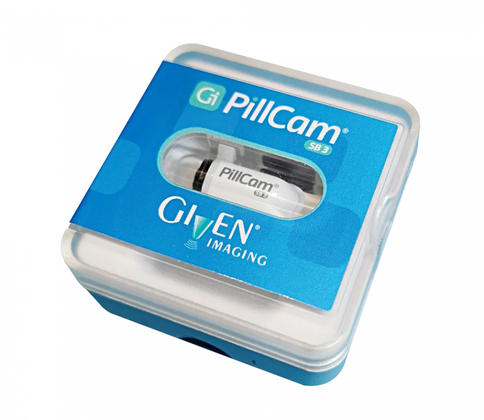
capsule endoscopy device
Biomarkers and targeted tests
Fecal calprotectin is a sensitive early marker; rising values can guide preemptive testing. Serologic panels, including CRP, and emerging tests for barrier proteins and microbial metabolites add detail. In some centers, analysis of mucosal microbiome and biofilm testing is becoming available, providing actionable information about persistent microbial communities.
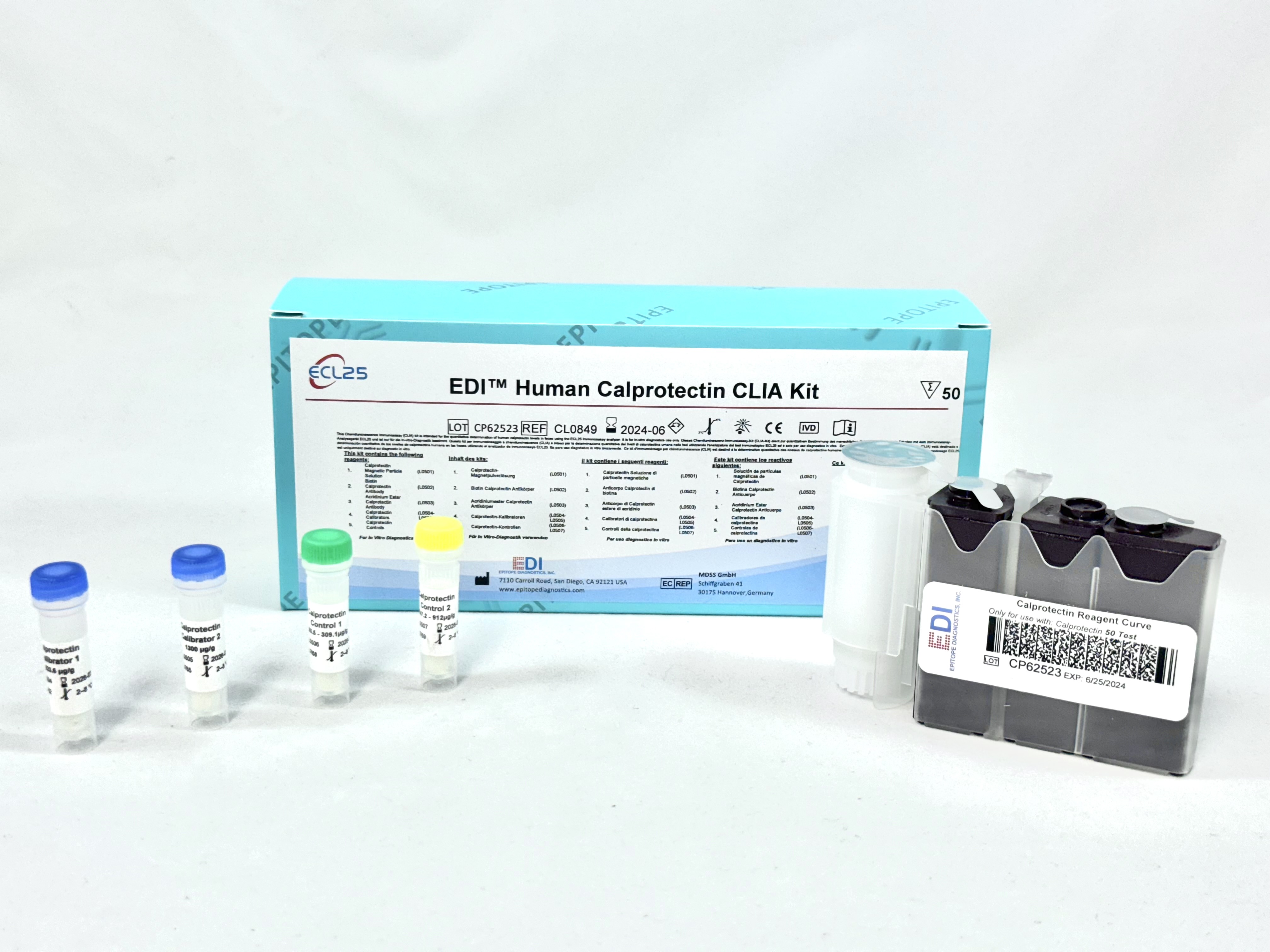
fecal calprotectin test kit
Treatment strategies for hidden defects
Treating these concealed contributors requires a layered, patient-specific plan that addresses anatomy, microbe, and immune response.
1. Surgical and endoscopic correction
Small strictures may respond to endoscopic balloon dilation or stricturoplasty, while persistent fistulas or diseased anastomoses sometimes require resection or revision. The point is judicious intervention: treating the structural source can reduce the need for escalating immunosuppression.
2. Microbial-targeted measures
Short, targeted antibiotic courses can suppress biofilm-related flares; adjuncts like anti-biofilm agents (where appropriate) and probiotics or microbiome restoration strategies may follow. Fecal microbiota transplant is experimental for Crohn’s but may have a role in selected ulcerative colitis cases and in clinical trials focused on refractory dysbiosis.
3. Optimizing medical therapy
Achieving deep remission—mucosal and preferably histologic—reduces recurrence risk. This may require optimizing biologic dosing, combining biologics with immunomodulators in specific settings, or choosing agents that better target the disease phenotype (e.g., anti-TNF for penetrating disease, anti-integrin for colonic disease). Personalized, biomarker-driven escalation and maintenance strategies are increasingly important.
Patient-centered monitoring and prevention
Long-term control demands active surveillance and shared decision-making. Establish a baseline with objective measures (calprotectin, endoscopy, imaging) and schedule surveillance based on disease type, surgery history, and risk factors. Smoking cessation, vaccination, stress management, and attention to diet and sleep are practical parts of reducing relapse triggers.
Real-world case vignette
A 34-year-old with ileal Crohn’s disease, with prior ileocolic resection, had several months of mild cramping and intermittent loose stools but normal CRP. Fecal calprotectin crept upward; MR enterography showed a subtle anastomotic narrowing with retained stool above it. Endoscopic dilation plus a short targeted antibiotic course and tightening of maintenance biologic dosing kept the patient in remission for years. The lesson: small anatomic problems, if found early, can be corrected with modest intervention.
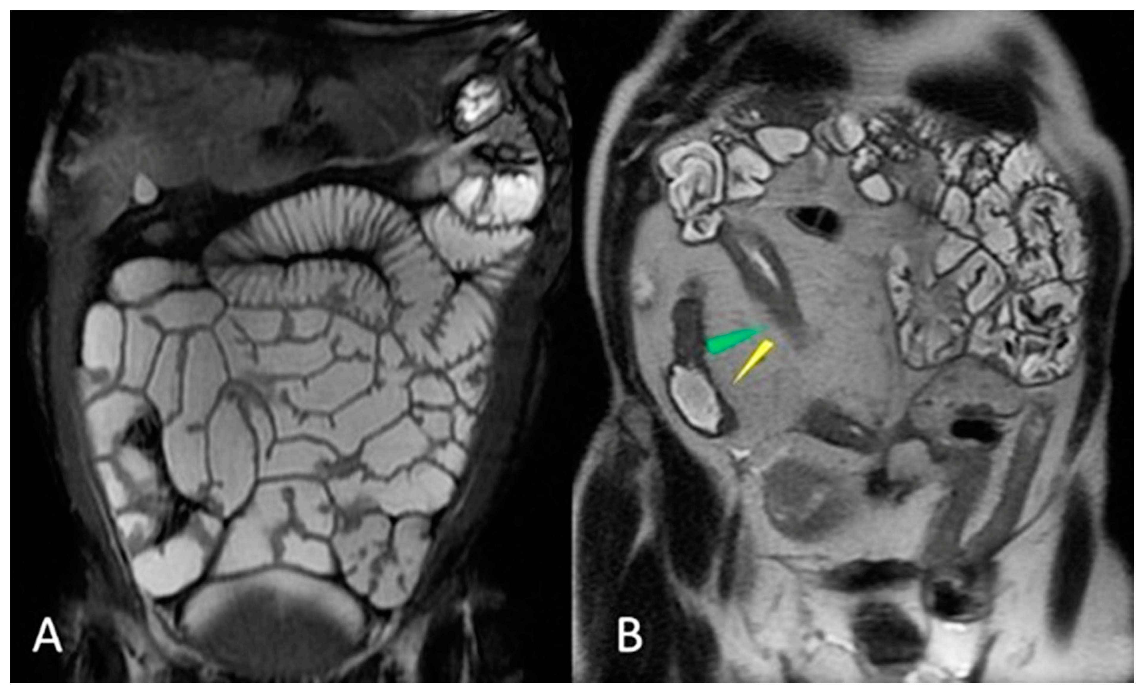
MR enterography small intestine
Living with uncertainty: practical advice for patients
IBD is a long game. Practically, that means cultivating partnerships with clinicians, tracking objective measures, and learning to recognize early signs of trouble. Keep a symptom log, request calprotectin tests when things feel off, and ask about advanced imaging if your history or prior surgery increases risk. Second opinions for persistent or unusual symptoms can be invaluable.
Where research is heading
Active research focuses on better imaging of biofilms, biomarkers that specifically reflect barrier integrity, and microbiome-editing therapies that go beyond blanket antibiotics. Precision medicine—using genomic, microbial, and immune profiling to predict who will relapse and what defect will cause it—is close to clinical reality and promises to make true durable remission attainable for many more patients.
Conclusion
For many with recurring IBD, the relapse story is not one of failed willpower or inevitable progression but of hidden anatomic and biological defects that quietly persist. Recognizing these issues—residual disease after surgery, strictures and blind loops, micro-fistulas, biofilm reservoirs, and incomplete mucosal healing—reframes the approach: look deeper, monitor smarter, and treat more precisely. When clinicians and patients adopt surveillance that seeks these concealed sources, relapses become less mysterious and more preventable.
- Recurrent IBD often stems from hidden structural or microbial defects, not just immune relapse.
- Early detection using fecal calprotectin, MR enterography, capsule endoscopy, and targeted endoscopy reduces the chance of full flares.
- Addressing anatomy (dilation, surgery), microbes (targeted antibiotics, microbiome strategies), and immune control together offers the best chance for durable remission.