
Full Clitoral Nerve Network Mapped: What This Means for Women's Health
The announcement that researchers have, for the first time, mapped the full clitoral nerve network marks a turning point in both medical science and public conversation about female sexual health. This mapping goes beyond a single anatomical sketch: it reveals an intricate web of sensory pathways, branching patterns, and connections to central nervous system hubs that were previously under-appreciated in clinical practice. The discovery challenges long-held assumptions, promises clearer treatment paths for chronic pelvic pain and sexual dysfunction, and forces medicine to recalibrate how it teaches, diagnoses, and performs surgery on female genital anatomy.
Why this mapping matters now
For decades, the clitoris and its nerve supply received far less scientific attention than male genital anatomy, a gap driven by cultural neglect and research priorities that often sidelined women's sexual experience. Accurate, high-resolution mapping addresses not only a scientific curiosity but also practical healthcare needs: better-informed surgical approaches, improved chronic pain management, and evidence-based sexual rehabilitation programs. Mapping the full nerve network validates the lived experiences of many women who reported sensory complexity that couldn't be explained by simplified diagrams.
"Seeing the clitoral nerve network in its full complexity reframes how we think about sensation, pain, and surgical risk in the pelvis."
A quick primer: clitoral anatomy and nerves
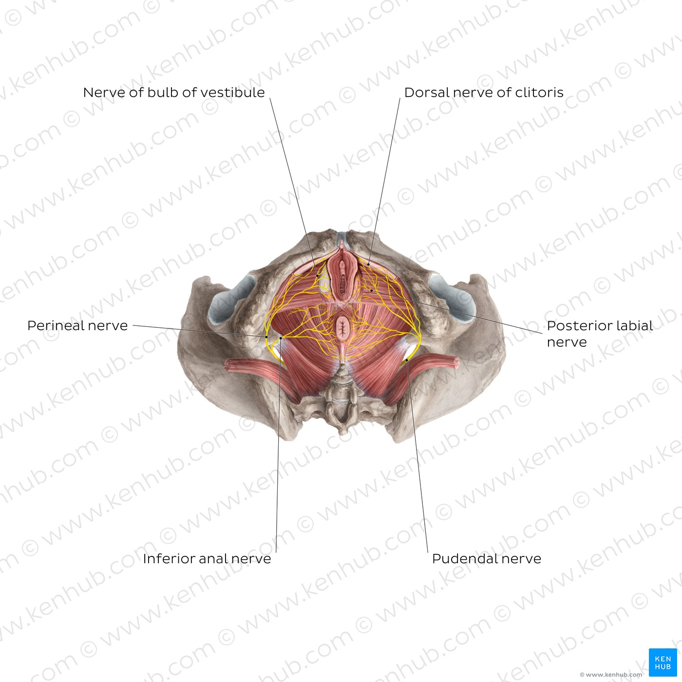
Before digging into the new map, it's helpful to summarize what clinicians have long accepted. The clitoris is more than an external glans: it includes internal crura and bulbs that extend along the pubic rami, tissues rich in specialized sensory receptors. Sensation travels from these receptors through peripheral nerves — historically described chiefly as branches of the dorsal nerve of the clitoris (a branch of the pudendal nerve) — toward the pelvic plexus and spinal cord. But that simplified account omitted smaller communicating branches and variations between individuals.

clitoral nerve anatomy diagram
How the mapping was done (methods explained)
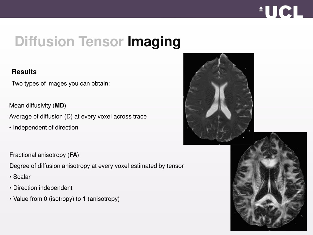
The full mapping combined modern imaging with meticulous dissection and neural tracing. Researchers used multimodal techniques: high-resolution MRI, diffusion tensor imaging (DTI) to infer fiber pathways, microdissection of cadaveric specimens to confirm branching, and histological staining to identify nerve fibers and receptor types. Some teams applied 3D reconstruction software to stitch together data from multiple specimens, revealing common patterns and anatomical variants.
These layered methods matter because any single approach has limits: imaging infers pathways but sometimes misses micro-branches; dissection reveals physical structures but cannot show living connectivity; histology confirms cell types but is inherently local. Together, they produced a robust, cross-validated map that clinicians can trust when planning interventions.
dorsal nerve of clitoris
Diagram: 3D reconstruction of clitoral sensory branches (schematic)
What the map revealed: unexpected complexity
Three findings stand out. First, the clitoral sensory network is much more distributed than the classic textbook model: multiple distinct nerve trunks and numerous microbranches reach the glans, hood, crura, and even adjacent labial tissues. Second, there are frequent anatomical variations — differences in branching patterns, crossover fibers, and connections with the pelvic plexus — that mean a "one-size-fits-all" surgical approach carries risk. Third, the map shows direct micro-connections between clitoral sensory fibers and other pelvic sensory pathways involved in pain modulation and autonomic regulation, offering a physiological explanation for why gynecologic or urologic conditions sometimes present with sexual dysfunction or altered sensation.
Clinical implications: surgery, pain, and sexual medicine
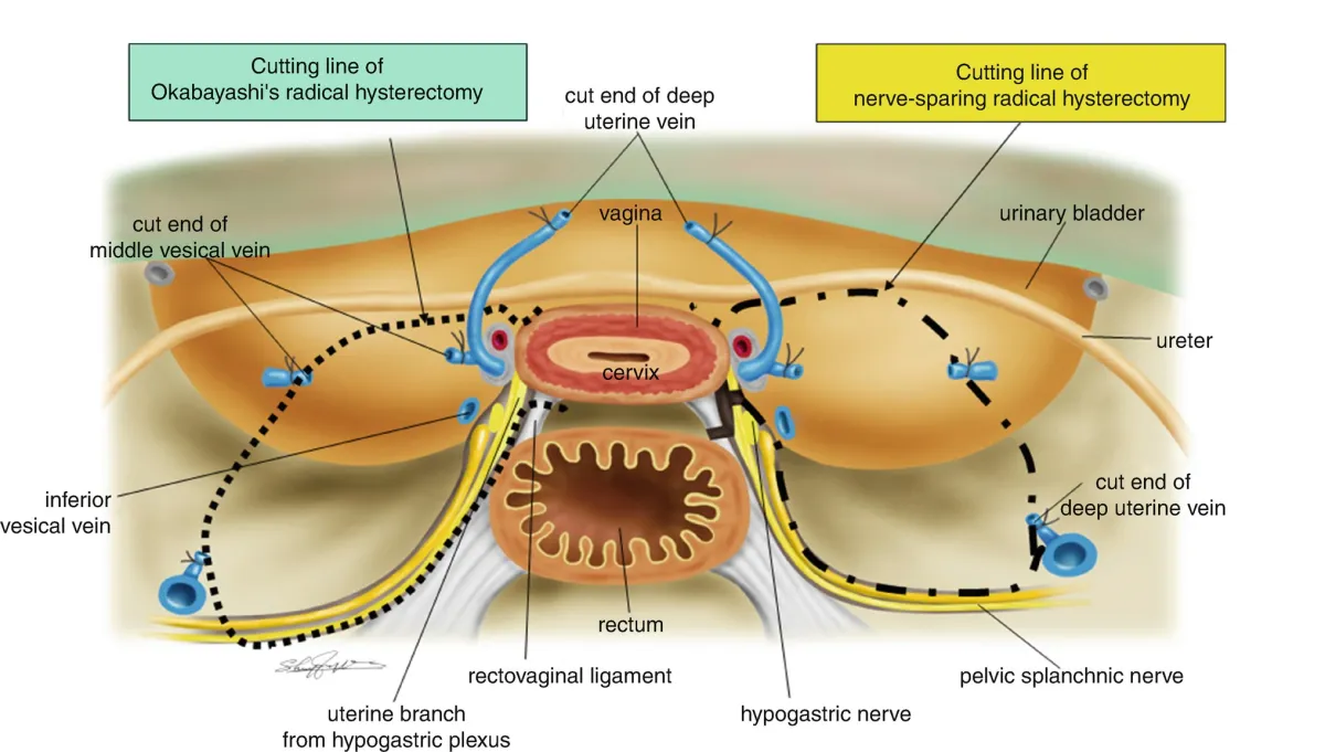
The map's practical impact is immediate. Surgeons performing pelvic or genital procedures — whether gender-affirming surgery, oncology resections, or routine gynecologic operations — can use the map to plan nerve-sparing approaches. Rather than relying on generalized anatomical landmarks, surgeons can anticipate common variants and avoid key trunks and branching zones to preserve sensation.
For patients with chronic pelvic pain or provoked vestibulodynia, the map offers diagnostic clarity. Pain that was once attributed solely to localized inflammation or psychosomatic factors may be traced to nerve entrapment, neuroma, or aberrant cross-talk between sensory pathways. That opens doors to targeted interventions: nerve blocks, microsurgical decompression, cryoneurolysis, neuromodulation, and multimodal rehabilitation that includes pelvic physical therapy and sexual counselling.

pelvic pain treatment nerve block
- Improved nerve-sparing surgical planning.
- Better-targeted pain treatments.
- Enhanced sexual rehabilitation protocols.
- More complex pre-op planning may increase imaging costs.
- Training required for surgeons and clinicians to use new maps effectively.
Implications for reconstructive and gender-affirming surgery
In reconstructive contexts — for example, after cancer surgery — knowing precisely where sensory trunks run allows microsurgeons to attempt nerve repairs or grafts with higher success probabilities. In gender-affirming genital surgeries, preserving or re-routing sensory nerves is paramount to achieving desired erotic and erogenous outcomes. The mapping supplies an anatomical foundation on which surgeons can standardize techniques, counsel patients more precisely about sensory outcomes, and develop new nerve-sparing protocols.

gender affirming genital surgery nerves
A new lens on sexual dysfunction and orgasm physiology
Sexual dysfunction is multifactorial, but the neural architecture matters. The full map suggests why different patients experience vastly different sensory profiles and why interventions such as pelvic-floor retraining, cognitive-behavioral therapy, or pharmacologic neuromodulation work for some women and not others. Clinicians can now consider whether a patient's complaints align with a peripheral neural issue, central sensitization, or a combination — and tailor treatment accordingly.
diffusion tensor imaging clitoris
"This map doesn't just redraw anatomy; it reorients clinical thinking about sensation as a dynamic network rather than a single pathway."
Ethical, educational, and policy considerations
Scientific breakthroughs in anatomy always carry ethical and social weight. First, medical curricula must be updated so that future physicians learn this complexity early, preventing inadvertent nerve injury across specialties. Second, patient education materials should reflect the realities of variation — informed consent must discuss not only common risks but also anatomical unpredictability and the limits of current predictive imaging.
There is also a justice dimension. Historically underfunded areas like female sexual health and pelvic pain have long suffered from delayed discovery. Policymakers and funders should view this mapping as evidence that investment yields actionable clinical advancements. Expanded coverage for diagnostic imaging, specialized pelvic pain clinics, and sexual rehabilitation services would translate anatomical knowledge into improved outcomes for patients.
nerve sparing hysterectomy technique
What this means for patients—real-world scenarios
Imagine a woman undergoing a hysterectomy who is concerned about postoperative changes in sexual sensation. With the new mapping, her surgical team can discuss specific nerve structures at risk and tailor the operative plan to minimize sensory loss. Or consider a patient with long-standing vulvar pain: rather than a long trial-and-error course of medications, a targeted nerve block informed by the map could provide rapid diagnostic and therapeutic benefit.
For survivors of genital trauma, the mapping offers pathways to sensory reconstruction and rehabilitation. Understanding nerve course and branching patterns allows microsurgeons and therapists to design staged approaches that combine surgical repair, graded sensory re-education, and psychosexual support.

vulvar pain neuromodulation
Limitations and unanswered questions
No single study answers every question. The current maps are based on combined imaging and cadaveric work and will need confirmation in diverse living populations across age, ethnicity, and medical history. How nerves adapt after injury, how peripheral mapping correlates with subjective sensation, and the best ways to measure sensory recovery remain active research areas. There are also technological hurdles: making high-resolution DTI and microstructural imaging widely accessible in clinical settings will require equipment, training, and protocols standardized across institutions.
Next steps for research and clinical translation
Researchers are likely to pursue several directions simultaneously: prospective clinical trials testing nerve-sparing surgical outcomes, controlled trials of targeted pain interventions guided by mapping, and longitudinal studies of sensory recovery after repair. On the technological side, improving in vivo imaging for routine preoperative assessment is a priority, as is incorporating anatomical variability into surgical simulation and training modules.
How clinicians should respond
Clinicians can begin by updating informed consent language and building multidisciplinary care paths that include pelvic pain specialists, pelvic floor physical therapists, sexual medicine clinicians, and mental health providers. Medical educators should integrate the new anatomic insights into anatomy labs, simulation training, and board exam preparation. Finally, hospitals and clinics should consider the value of investing in diagnostic imaging and training that directly reduce complication rates and improve patient-reported outcomes.
Conclusion: a foundational advance with cascading benefits
Mapping the full clitoral nerve network is more than a technical achievement: it is a corrective to the history of under-recognized female anatomy, and a practical tool that will change surgical practice, pain medicine, sexual rehabilitation, and medical education. The immediate beneficiaries are patients who will receive clearer diagnoses and more targeted treatments. The longer-term payoff will be a shift in how medicine thinks about genital sensation — not as a single line from point A to point B, but as a network whose preservation requires knowledge, care, and humility.
- Researchers have produced the first comprehensive map of the clitoral nerve network, revealing complex branching and anatomical variability.
- The map supports nerve-sparing surgical techniques, more precise pain interventions, and improved sexual medicine practice.
- Implementation requires updates to clinical consent, training, imaging access, and multidisciplinary care pathways.
Final thought
Scientific progress often arrives incrementally; occasionally it redraws the map. This mapping does the latter for female genital anatomy—opening new clinical possibilities and, perhaps more importantly, inviting a medical culture that listens more closely to women's experiences and treats them with the anatomical respect they deserve.