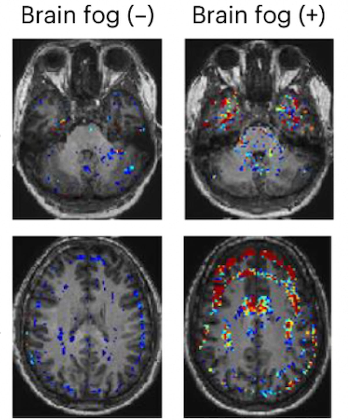
Brain Scans Suggest Vascular Cause for Long COVID Brain Fog
When people describe long COVID as a fog that settles over thought and stamina, they are giving language to something millions experience but scientists and clinicians have struggled to explain: persistent, disabling fatigue and cognitive dysfunction that follow SARS-CoV-2 infection. In the past few years a growing body of neuroimaging work — from glucose PET studies to cerebral blood-flow MRI — has revealed reproducible patterns in patients with post‑acute symptoms. Those patterns point to one plausible, and testable, unifying idea: microvascular and perfusion disturbances in the brain may underlie at least some cases of long COVID fatigue and brain fog.
neuroimaging long COVID brain
What researchers are seeing in scans
PET: A recurring pattern of hypometabolism
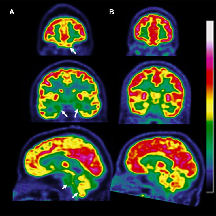
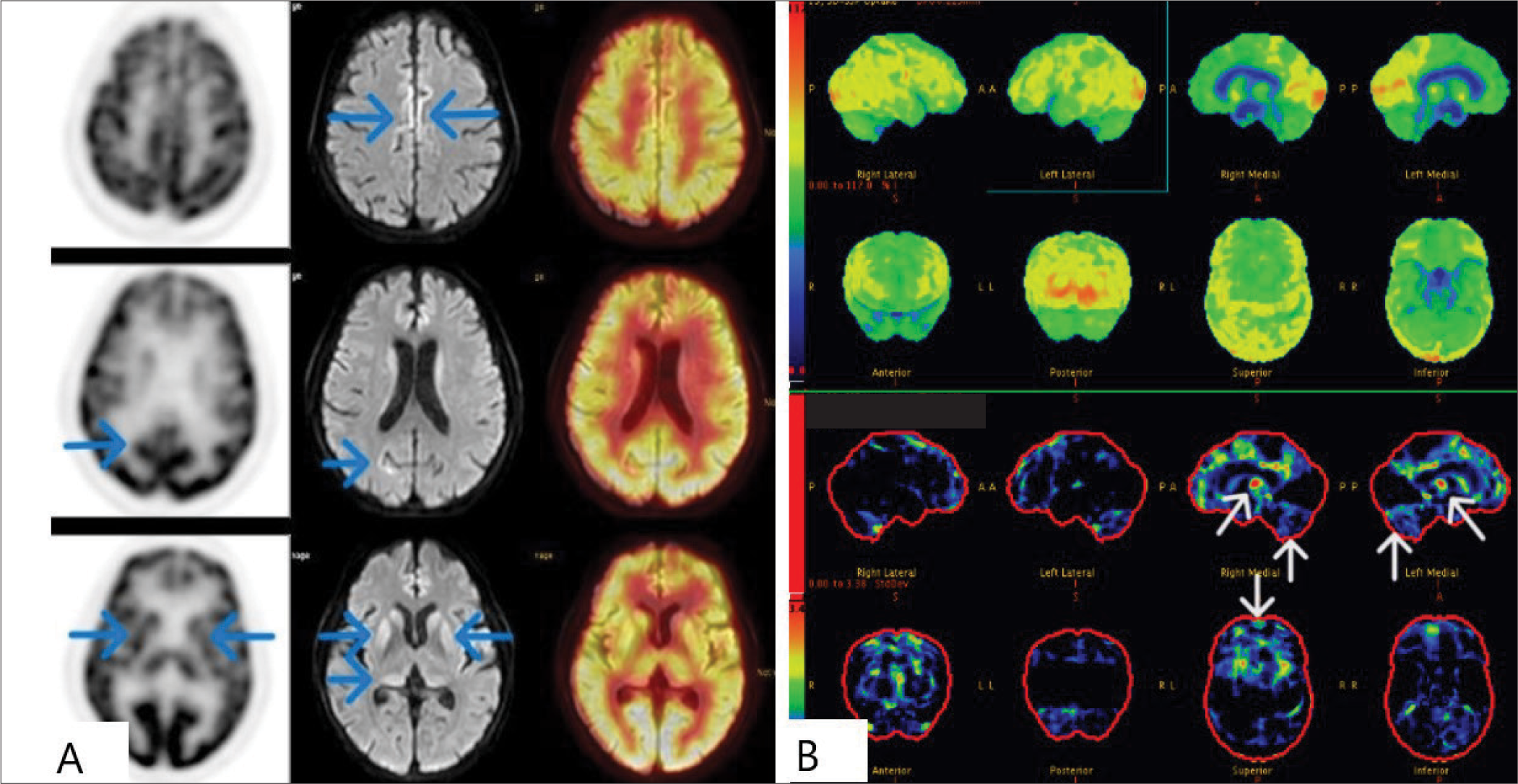
Multiple teams using 18F‑FDG PET — which images glucose metabolism as a proxy for neuronal and synaptic activity — have reported similar spatial patterns of reduced metabolism in people with persistent post‑COVID symptoms. The affected regions commonly include orbitofrontal and olfactory-related areas, limbic and paralimbic structures, parts of the frontal and temporal lobes, and in many studies the brainstem and cerebellum as well. These metabolic reductions track with symptom burden: patients with worse fatigue, memory complaints, or more symptoms during acute infection tend to show more extensive hypometabolism on PET. citeturn0search1
FDG-PET brain hypometabolism
MRI and perfusion: decreased cerebral blood flow
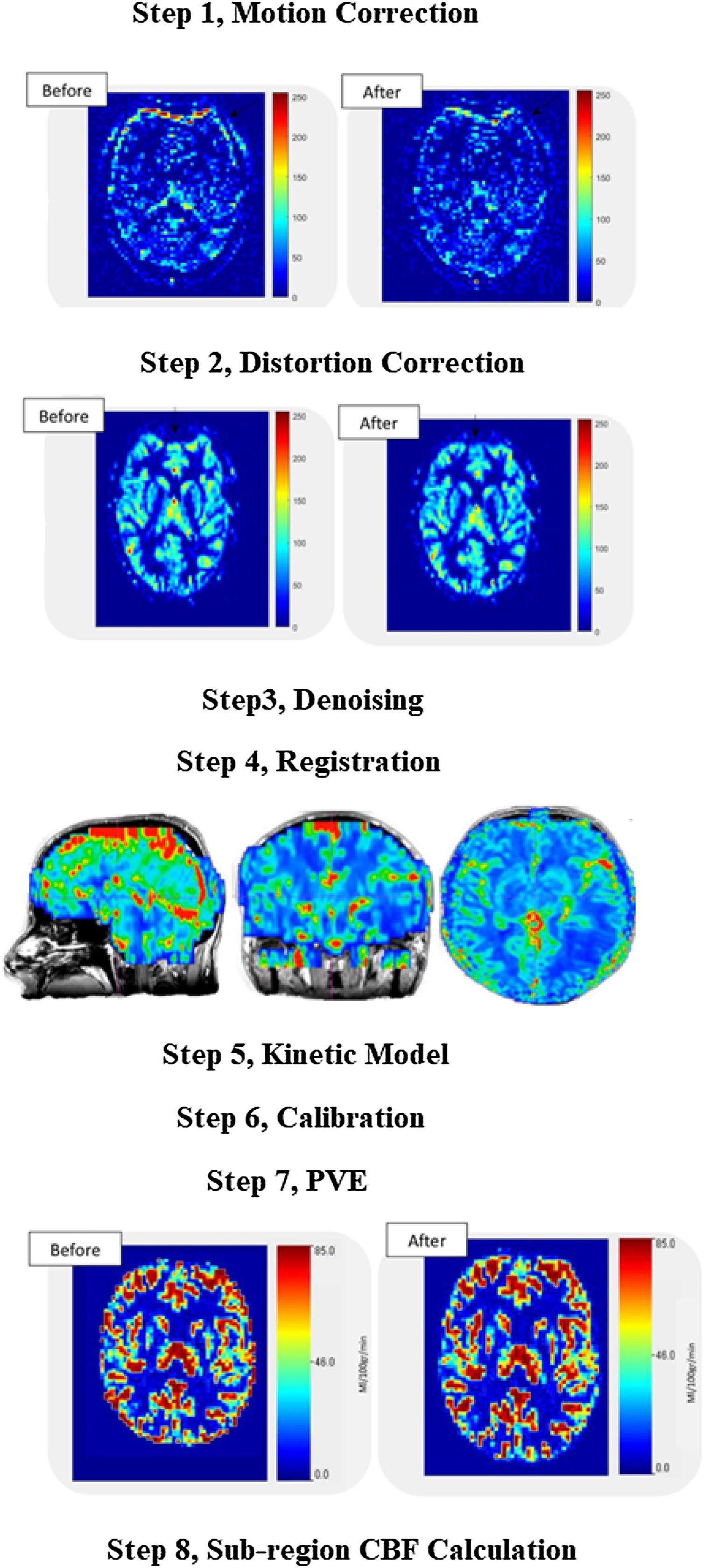
Arterial spin labeling (ASL) MRI and other perfusion-sensitive sequences have independently found reductions in cerebral blood flow (CBF) in cohorts of recovered COVID‑19 patients, even those who were never hospitalized. Lower CBF in specific regions has been correlated with higher fatigue scores and poorer immediate memory performance on neuropsychological tests, suggesting a direct link between perfusion deficits and clinical symptoms. citeturn0search9
ASL MRI cerebral blood flow
Time course and variability
A further important observation is that these imaging abnormalities are not identical across every patient and they may change over time. Some studies show partial recovery of metabolic patterns over months in some people, while others report persistent changes beyond six months. The trajectories appear heterogeneous, which hints at multiple interacting mechanisms — some reversible, others possibly longer lasting. citeturn0search6
"The scans don't prove a single cause, but they consistently point to impaired brain energy use and blood supply as central players in the experience of long COVID."
How microvascular problems could explain fatigue and brain fog
Why perfusion matters to cognition and stamina
Neurons and the cells that support them are extraordinarily metabolically active: they need a steady supply of oxygen and glucose, and they rely on tightly regulated microcirculation at the capillary level. Small changes in local perfusion can disrupt synaptic signaling, slow processing speed, and reduce the brain's ability to sustain prolonged mental or physical effort. In plain terms, when capillaries don't deliver enough oxygen or glucose to circuits involved in attention, executive function, and arousal, subjective fatigue and cognitive slowing follow.
Microvascular injury and capillary loss
Pathology and animal-model studies of SARS‑CoV‑2 have documented microvascular damage in the brain, including signs of endothelial cell injury and loss of capillary integrity. Some autopsy work has described 'string vessels' — remnants of capillaries that have lost their endothelial lining — and other microvascular changes that would be expected to reduce effective perfusion at a tissue level. These findings provide a plausible anatomical substrate for the perfusion and metabolic signals seen on PET and ASL MRI. citeturn1search0turn1search1
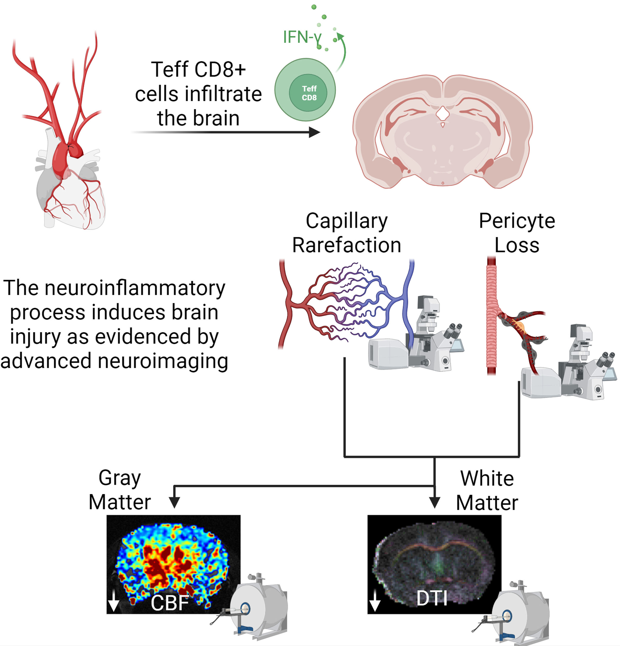
microvascular injury brain capillaries
Microclots and capillary blockage
Another mechanism receiving attention is the formation of micro‑scale, fibrin‑rich aggregates (often called fibrinaloid microclots) that resist normal breakdown. The hypothesis is that these microscopic obstructions can lodge in capillary beds and impair oxygen delivery without producing the large-vessel strokes doctors traditionally look for. Several groups have reported higher microclot burden and abnormal coagulation-related biomarkers in long COVID cohorts; if present in cerebral microcirculation, such clots could translate into diffuse, low‑grade ischemia and the scan abnormalities clinicians observe. citeturn1search2
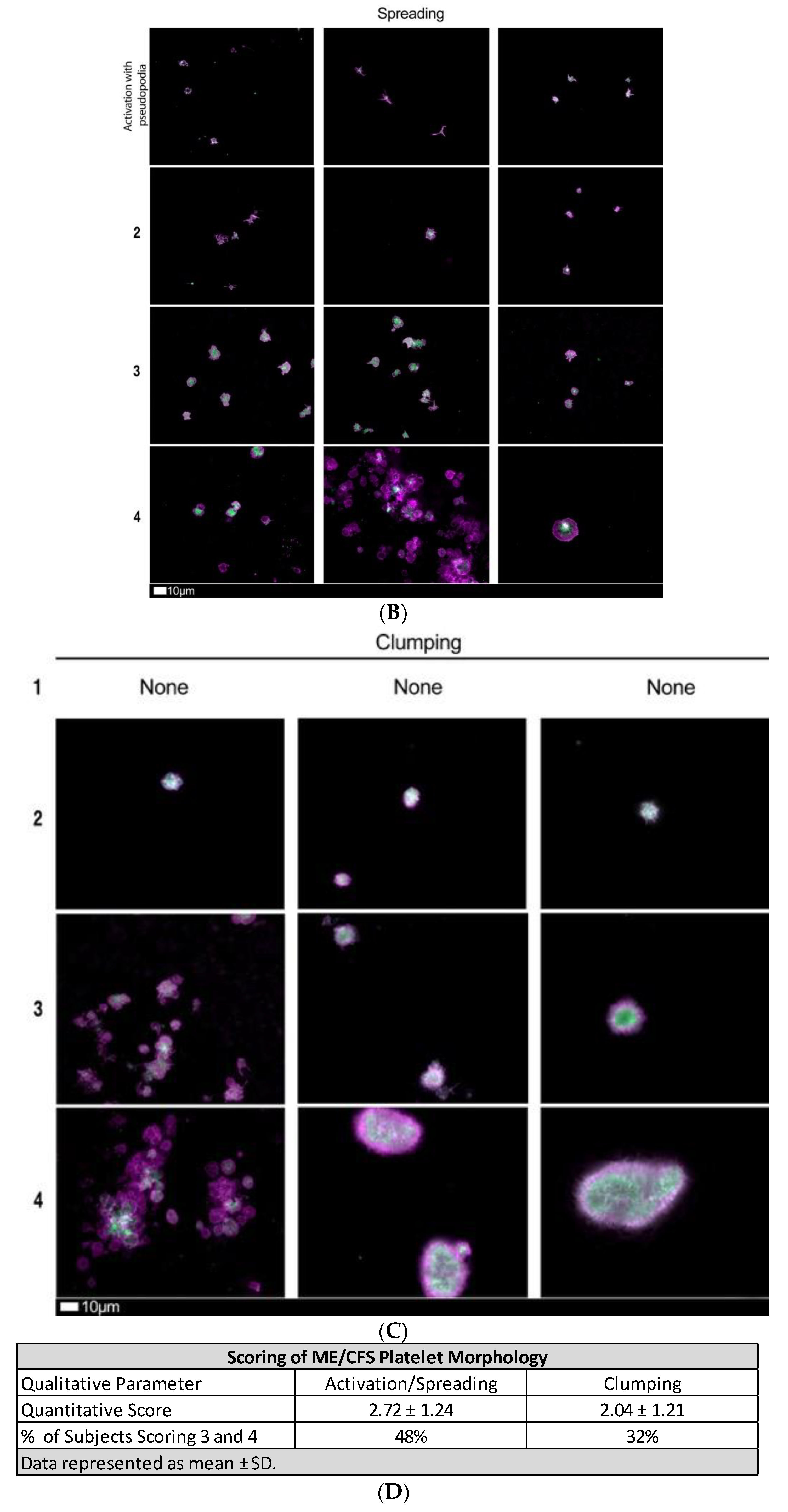
fibrinaloid microclots microscopy
Neuroinflammation and endothelial dysfunction
Inflammation, whether systemic or localized within the brain, can alter endothelial cell function, promote coagulation, and disrupt the blood‑brain barrier. Microglial and astrocyte activation have been reported in neuropathology studies of COVID‑19, and inflammatory signaling can impair neurovascular coupling — the process that matches blood flow to neuronal activity. When neurovascular coupling falters, the brain's metabolic demands are not met efficiently; PET and CBF signatures will reflect that mismatch even in the absence of frank tissue loss. citeturn1search11
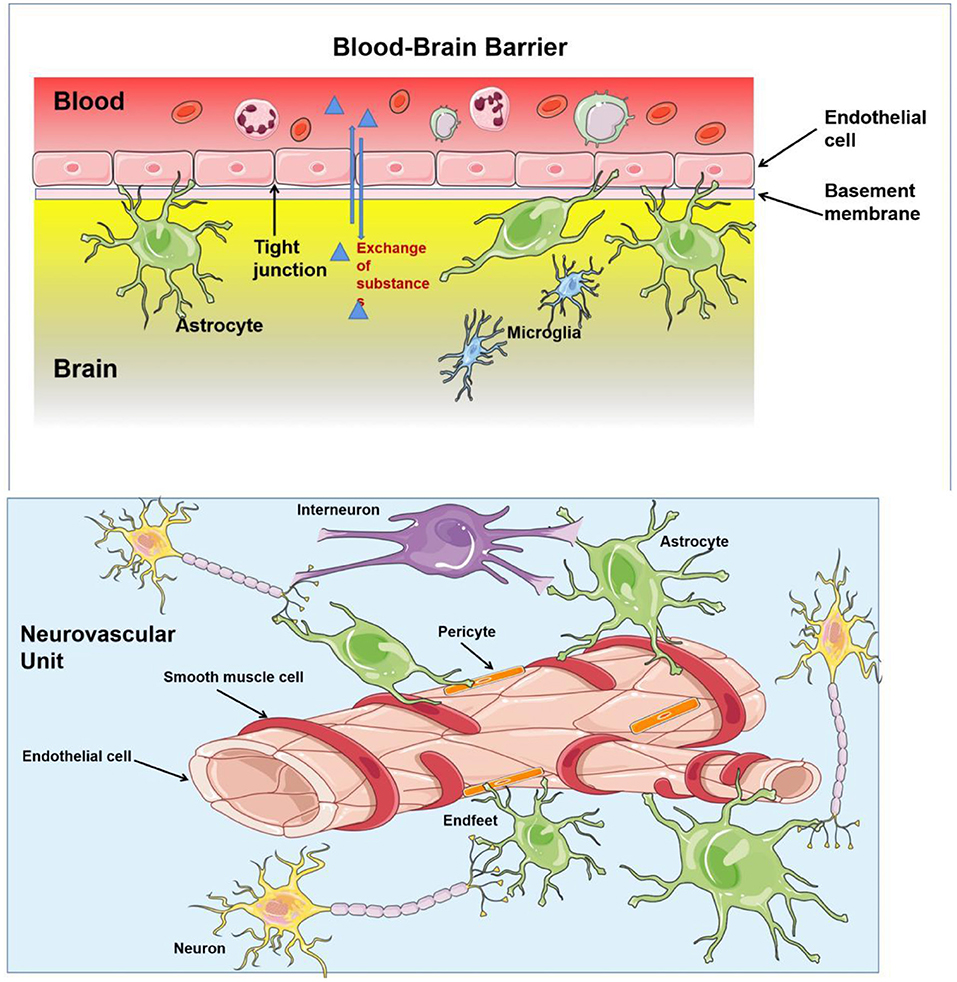
brain endothelial dysfunction
Putting the pieces together: an integrated model
Viewed together, these data support an integrative model in which one or more of the following — endothelial injury from viral or immune-mediated mechanisms, thrombotic microclots, persistent inflammation, and disrupted neurovascular signaling — reduce microvascular efficiency and regional perfusion. Reduced perfusion then produces measurable hypometabolism on PET, which aligns with the clinical picture of fatigue, post‑exertional malaise, and cognitive slowing. This is not a single-pathway story for all patients; instead, it's a framework that can accommodate heterogeneity while suggesting concrete targets for diagnosis and intervention.
Clinical implications: diagnosis, monitoring, and trials
Imaging as a biomarker
Brain FDG‑PET and ASL MRI are emerging as candidate biomarkers for objective assessment of brain involvement in long COVID. In clinical research settings they can help stratify patients, correlate symptoms with physiology, and measure response to interventions — for example, drugs that target inflammation, endothelial health, or coagulation. Importantly, these tools are not yet standard-of-care for long COVID and access is limited; PET in particular is expensive and not widely available for routine diagnostic use.
Therapeutic strategies under investigation
Because the proposed mechanisms span vascular, inflammatory, and coagulation pathways, multiple therapeutic strategies are being explored: anti‑inflammatory and immunomodulatory agents, therapies aimed at improving endothelial function, and anticoagulant or antiplatelet regimens designed to reduce microclot burden. A few small, early reports and preprints suggest symptomatic improvement in some patients after targeted antithrombotic therapy, but larger randomized trials are necessary to determine safety and efficacy. At present, most treatments remain experimental and should only be pursued in the context of clinical studies or after specialist consultation. citeturn1search3turn1search14
Rehabilitation and pragmatic care
While researchers test pharmacological approaches, clinicians recommend practical, evidence‑based measures: graded return-to-activity with attention to post‑exertional symptom exacerbation, cognitive rehabilitation strategies, sleep optimization, and treatment of comorbid contributors such as orthostatic intolerance or mood disorders. Individualized care plans that respect the variable trajectory of recovery remain the mainstay for now.
Limitations, uncertainties, and unanswered questions
Several important caveats must temper enthusiasm. First, correlation on scans does not prove causation; the observed hypometabolism and perfusion changes could be downstream effects of other systemic problems. Second, many imaging studies so far have relatively small sample sizes, variable timing from acute infection to imaging, and differing inclusion criteria — factors that complicate comparisons. Third, the microclot literature includes promising but not yet definitive human trial evidence, and measurement techniques for fibrinaloid microclots remain variable across labs. Finally, individual vulnerability — including pre‑existing vascular risk factors, age, and genetic contributors — likely modifies risk and recovery, and we still lack reliable predictors of who will develop persistent symptoms. citeturn0search4turn1search6
What patients should know
For people living with long COVID, the emerging imaging findings are both validating and cautiously hopeful. They provide objective evidence that the symptoms are real and tied to measurable changes in brain physiology. At the same time, the science is evolving: treatments that correct the underlying biology remain under study, and recovery is variable. Patients should seek multidisciplinary care, prioritize symptom management and pacing, and consider participation in research studies that help expand understanding and access to new therapies.
Conclusion: where research goes next
Neuroimaging has moved long COVID from anecdote toward anatomy. PET and perfusion MRI converge on a story of altered metabolism and blood flow that plausibly links microvascular dysfunction to fatigue and brain fog. The next steps are clear: larger longitudinal imaging cohorts, standardized microclot assays, randomized trials of targeted vascular and anti‑inflammatory therapies, and integrative studies that combine imaging, blood biomarkers, and cognitive testing. If microvascular dysfunction proves to be a dominant mechanism in a substantial subset of patients, it opens a pathway to precision diagnostics and mechanistically guided treatments — a shift from managing symptoms to treating a biological cause.
- Consistent imaging signals: FDG‑PET and perfusion MRI repeatedly show hypometabolism and reduced blood flow in many people with long COVID.
- Microvascular hypothesis: Endothelial injury, capillary loss, and microclots are plausible mechanisms that could impair brain perfusion and energy use.
- Clinical translation: Imaging may help stratify patients and guide trials, but routine clinical use is not yet established.
- Research priorities: Larger longitudinal studies and randomized trials of vascular or anticoagulant strategies are needed.
Acknowledgment
This article synthesizes current neuroimaging and pathophysiological evidence to offer a cohesive explanation for long COVID fatigue and brain fog; the field is advancing rapidly and readers should consult specialized clinics and emerging clinical trials for the latest guidance.